. Scientific Frontline

Wednesday, August 31, 2022

Study finds tiny brain area controls work for rewards

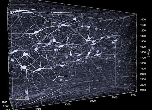
The lateral habenula in the mouse brain, with axons streaming down to dopaminergic and serotonergic centers. Credit: Warden Lab

A tiny but important area in the middle of the brain acts as a switch that determines when an animal is willing to work for a reward and when it stops working, according to a study published Aug. 31 in the journal Current Biology.

“The study changes how we think about this particular brain region,” said senior author Melissa Warden, assistant professor and Miriam M. Salpeter Fellow in the Department of Neurobiology and Behavior, which is shared between the College of Arts and Sciences and the College of Agriculture and Life Sciences.

“It has implications for psychiatric disorders, particularly depression and anxiety,” Warden said.

The paper, “Tonic Activity in Lateral Habenula Neurons Acts as a Neutral Valence Brake on Reward-Seeking Behavior,” illuminates the role of the lateral habenula, a small structure on top of the thalamus, which funnels higher-level information from the front and center of the brain to areas that produce neurotransmitters such as serotonin and dopamine.

The lateral habenula’s exact role has been unclear until now. The new study shows that when neurons in this brain area turn off, an animal will work for rewards; when those neurons fire, the animal becomes disengaged and stops working. Experiments revealed that the lateral habenula turns on specifically when an animal has had enough of a reward and is satisfied, or when it finds its work no longer yields a reward.

Signs of Saturation Emerge from Particle Collisions at RHIC

Brookhaven Lab physicists Xiaoxuan Chu and Elke-Caroline Aschenauer at the STAR detector of the Relativistic Heavy Ion Collider (RHIC).
Source/Credit: Brookhaven National Laboratory

Nuclear physicists studying particle collisions at the Relativistic Heavy Ion Collider (RHIC)—a U.S. Department of Energy Office of Science user facility at DOE’s Brookhaven National Laboratory—have new evidence that particles called gluons reach a steady “saturated” state inside the speeding ions. The evidence is suppression of back-to-back pairs of particles emerging from collisions between protons and heavier ions (the nuclei of atoms), as tracked by RHIC’s STAR detector. In a paper just published in Physical Review Letters, the STAR collaboration shows that the bigger the nucleus the proton collides with, the larger the suppression in this key signature, as predicted by theoretical models of gluon saturation.

“We varied the species of the colliding ion beam because theorists predicted that this sign of saturation would be easier to observe in heavier nuclei,” explained Brookhaven Lab physicist Xiaoxuan Chu, a member of the STAR collaboration who led the analysis. “The good thing is RHIC, the world’s most flexible collider, can accelerate different species of ion beams. In our analysis, we used collisions of protons with other protons, aluminum, and gold.”

Peering Into Mirror Nuclei

Diagram showing a high-energy electron scattering from a correlated nucleon in the mirror nuclei tritium (left) and helium-3 (right). The electron exchanges a virtual photon with one of the two correlated nucleons, knocking it out of the nucleus and allowing its energetic partner to escape. Both nuclei have neutron-proton pairs, while tritium has an additional neutron pair and helium-3 has an additional proton pair.
Credit: Jenny Nuss/Berkeley Lab

The atomic nucleus is a busy place. Its constituent protons and neutrons occasionally collide, and briefly fly apart with high momentum before snapping back together like the two ends of a stretched rubber band. Using a new technique, physicists studying these energetic collisions in light nuclei found something surprising: protons collide with their fellow protons and neutrons with their fellow neutrons more often than expected.

The discovery was made by an international team of scientists that includes researchers from the Department of Energy’s Lawrence Berkeley National Laboratory (Berkeley Lab), using the Continuous Electron Beam Accelerator Facility at DOE’s Thomas Jefferson National Accelerator Facility (Jefferson Lab) in Virginia. It was reported in a paper published today in the journal Nature.

Understanding these collisions is important for interpreting data in a wide range of physics experiments studying elementary particles. It will also help physicists better understand the structure of neutron stars – collapsed cores of giant stars that are among the densest forms of matter in the universe.

John Arrington, a Berkeley Lab scientist, is one of four spokespersons for the collaboration, and Shujie Li, the lead author on the paper, is a Berkeley Lab postdoc. Both are in Berkeley Lab’s Nuclear Science Division.

"Greener" Fertilizer and Carbon-free Fuels Come Closer to Reality

Photo by Richard Bell on Unsplash

A little over 100 years ago, humankind learned how to take nitrogen from the atmosphere (where it is plentiful) and turn it into ammonia that can be used as source of fertilizer for growing food. That chemical process, known as nitrogen fixation, has allowed huge increases in crop production and a subsequent boom in human populations fed by those crops.

Nearly all artificial nitrogen fixation is done with what is known as the Haber–Bosch process, which uses a metal catalyst to combine gaseous nitrogen and hydrogen into ammonia, at high pressures and temperatures. Ammonia fixed through this process is estimated to be responsible for growing crops that feed half the world's population.

But there is another large source of nitrogen fixation: bacteria that live in soil, which fix nitrogen at normal atmospheric temperatures and pressures. In recent decades, researchers searching for sustainable agriculture practices have looked to these microbes as inspiration for developing nitrogen-fixation processes that are easier to conduct and more environmentally friendly than the energy-intensive Haber-Bosch process. Now, a team at Caltech led by Jonas Peters, Bren Professor of Chemistry and director of the Resnick Sustainability Institute, has made a breakthrough that increases the efficiency of one of these low-temperature and low-pressure processes, further opening the door to greener fertilizer, and even the production of zero-carbon fuels.

Climate change and ocean oxygen

Oxygen-deficient zones (in red) shrank during long warm periods in the past, contrary to widespread expectations.   
Image Credit: Alexandra Auderset, Princeton and MPIC

In the last 50 years, oxygen-deficient zones in the open ocean have increased. Scientists have attributed this development to rising global temperatures: Less oxygen dissolves in warmer water, and the tropical ocean’s layers can become more stratified.

But now, contrary to widespread expectations, an international team of scientists led by researchers from the Max Planck Institute for Chemistry and Princeton University has discovered that oxygen-deficient zones shrank during long warm periods in the past.

“We had not expected such a clear effect,” said Alexandra Auderset, first author of the new paper in the journal Nature and currently a visiting postdoctoral research fellow at Princeton University. She led the study with Alfredo Martínez-García at the Max Planck Institute for Chemistry in Mainz, as part of a long-term collaboration with Daniel Sigman’s group at Princeton University.

Understanding these changes is important because “when oxygen becomes scarce, life has a harder time,” said Sigman, Dusenbury Professor of Geological and Geophysical Sciences. For example, in low-oxygen regions of the eastern Pacific and northern Indian Ocean, only specialized microbes and organisms with a slow metabolism — such as jellyfish — can survive.

Marine Protected Areas in Antarctica should include young emperor penguins, scientists say

A group of Juvenile emperor penguins at Atka Bay on the sea ice edge ready for their first swim. In four years, they will return to breed, spending much of their time in unprotected areas of the Southern Ocean.
Image credit: Daniel P. Zitterbart/ ©Woods Hole Oceanographic Institution

Scientists at the Woods Hole Oceanographic Institution (WHOI) and European research institutions are calling for better protections for juvenile emperor penguins, as the U.S. Fish and Wildlife Service considers listing the species under the Endangered Species Act and the Commission for the Conservation of Antarctic Marine Living Resources (CCAMLR) considers expanding the network of Marine Protected Areas (MPAs) in the Southern Ocean.

In one of the few long-term studies of juvenile emperor penguins–and the only study focused on a colony on the Weddell Sea–research published today in Royal Society Open Science found that the young birds spend about 90 percent of their time outside of current and proposed MPAs. The study, which tracked eight penguins with satellite tags over a year, also found that they commonly traveled over 1,200 kilometers (745 miles) beyond the species range defined by the International Union for Conservation of Nature (IUCN), which is based on studies of adult emperor penguins from a few other colonies.

Considered immature until about 4 years of age, juvenile emperor penguins are more vulnerable than adults because they have not fully developed foraging and predator avoidance skills. As climate change reduces sea-ice habitat and opens up new areas of the Southern Ocean to commercial fishing, the researchers conclude that greatly expanded MPAs are crucial to protect this iconic, yet threatened, penguin species at every life stage.

Brain activity during sleep differs in young people with genetic risk of psychiatric disorders

Photo by Lux Graves on Unsplash

Young people living with a genetic alteration that increases the risk of psychiatric disorders have markedly different brain activity during sleep, a study led by researchers from the Universities of Bristol and Cardiff published in the journal eLife shows.

The brain activity patterns during sleep shed light on the neurobiology behind a genetic condition called 22q11.2 Deletion Syndrome (22q11.2DS) and could be used as a biomarker to detect the onset of neuropsychiatric disorders in people with 22q11.2DS.

Caused by a gene deletion of around 30 genes on chromosome 22, 22q11.2DS occurs in one in 3000 births. It increases the risk of intellectual disability, autism spectrum disorder (ASD), attention-deficit hyperactivity disorder (ADHD) and epileptic seizures. It is also one of the largest biological risk factors for schizophrenia. However, the biological mechanisms underlying psychiatric symptoms in 22q11.2DS are unclear.

Marianne van den Bree, co-senior author and Professor of Psychological Medicine at Cardiff said: “We have recently shown that the majority of young people with 22q11.2DS have sleep problems, particularly insomnia and sleep fragmentation, that are linked with psychiatric disorders. However, our previous analysis was based on parents reporting on sleep quality of their children, and the neurophysiology – what’s happening to brain activity – has not yet been explored.”

Scientists Determined Content of Harmful Substances in Ekaterinburg Mud

In Ekaterinburg, scientists made more than 60 mud samples.
Photo credit: Ilya Safarov

Scientists at Ural Federal University and the Institute of Industrial Ecology Ural Branch of the Russian Academy of Sciences, together with their colleagues from Southern Federal University, Egypt and Saudi Arabia, have studied the concentration of potentially harmful substances in surface sediments in Ekaterinburg and identified possible sources of pollution. The research contributes to the development and adoption of standards for the content of harmful substances in mud. The results of the research, funded by the Russian Science Foundation (Project №18-77-10024), are described in the journal Chemosphere.

Over several seasons, scientists took more than 60 mud samples collected at the same sites: first, in green areas with flower beds and lawns, second, from roads and, third, from neighborhoods, parking lots and sidewalks in residential areas of the 1.5 million metropolises. Dirt samples were dried under laboratory conditions for several weeks and then passed through a 1-mm sieve. The resulting dust was analyzed.

"In samples from all surveyed areas the highest concentrations of iron and manganese are found, as they are contained in rocks and soils, on which Yekaterinburg is located. Dust from green areas, in addition, has high concentrations of zinc and lead, in samples from roads - cobalt, nickel, tin and antimony, dust from yard passages, parking lots and sidewalks is full of zinc and copper," says Andrian Seleznev, Associate Professor of Department of Health and Safety at UrFU, Senior Scientist of Industrial Ecology Ural Branch of the Russian Academy of Sciences.

Archaeology and ecology combined sketch a fuller picture of past human-nature relationships

Hunting of a deer. Wall painting, 6th millennium BC. Museum of Anatolian Civilizations, Ankara.
 Image source: Wikimedia Commons

For decades now, archaeologists wielded the tools of their trade to unearth clues about past peoples, while ecologists have sought to understand current ecosystems. But these well-established scientific disciplines tend to neglect the important question of how humans and nature interacted and shaped each other across different places and through time. An emerging field called archaeoecology can fill that knowledge gap and offer insights into how to solve today’s sustainability challenges, but first, it must be clearly defined. A new paper by SFI Complexity Fellow Stefani Crabtree and Jennifer Dunne, SFI’s Vice President for Science, lays out the first comprehensive definition of archaeoecology and calls for more research in this nascent but important field.

While an archaeology or palaeobiology study might examine a particular relationship, such as how humans in New Guinea raised cassowaries during the Late Pleistocene, archaeoecology takes a much broader view. “It’s about understanding the whole ecological context, rather than focusing on one or two species,” Dunne explains.

3D imaging contributes to a better understanding of early stages of Alzheimer's disease

Three-dimensional image of noradrenergic nerve cells in the envelope of locus coeruleus.
Photo credit: Gilvesy et al.

With the help of a new imaging technique for 3D, researchers at Karolinska Institutet, among others, have been able to characterize a part of the brain that shows the most accumulation of tau protein, an important biomarker for the development of Alzheimer's disease. The results published in the journal Acta Neuropathologica may in the future make it possible to have a more accurate neuropathological diagnosis of Alzheimer's disease spectrum at a very early stage.

Intracellular accumulation of pathological tau protein in the brain is a hallmark of several age-related neurodegenerative diseases, including Alzheimer's disease, which accounts for 60-80 percent of all dementia cases worldwide.

In a new study, researchers at Karolinska Institutet, SciLifeLab in Stockholm and several universities from Hungary, Canada, Germany and France have applied a state-of-the-art immune imaging technology, in combination with light sheet microscopy, to investigate a human brain stem core, locus coeruleus, which is an important core in the mammalian brain.

Featured Article

Autism and ADHD are linked to disturbed gut flora very early in life

The researchers have found links between the gut flora in babies first year of life and future diagnoses. Photo Credit:  Cheryl Holt Disturb...

Top Viewed Articles