. Scientific Frontline

Thursday, October 28, 2021

Engineering molecules to turn off inflammation

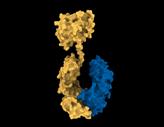
Stylized scientific image of the anti-inflammatory
molecule, Interleukin-37
In a long-term collaboration between researchers and industry, an exciting first step has been made in the creation of a new generation of medicines for auto-immune diseases such as arthritis and inflammatory bowel disease using one of our body’s own anti-inflammatory off-switch molecules.

Our body’s own immune system produces many highly potent anti-inflammatory molecules, but they are often highly fragile, short-lived, and do not have drug-like properties. Interleukin-37 is one such molecule produced by the body to turn off inflammation.

Together with partner F. Hoffmann-La Roche (Roche), the multidisciplinary research team from the Monash University Biomedicine Discovery (BDI) Institute, Monash University’s Department of Pediatrics and the Hudson Institute of Medical Research have harnessed their Fc-fusion platform to engineer the next generation of Interleukin-37, one that retains anti-inflammatory potency, is highly stable and has an excellent therapeutic likeness.

The findings from the research collaboration have now been published in Cell Chemical Biology.

A little bit of inflammation can be a good thing and is often the body's immune system doing its job. However, when inflammation persists, or the immune system starts attacking the body’s own cells, this can lead to disease.

One of the study’s lead authors Dr. Andrew Ellisdon from the Monash BDI says many human diseases, including autoimmune diseases such as arthritis or inflammatory bowel disease, are characterized by too much inflammation. There is a gap in producing new generations of potent anti-inflammatory therapeutics.

Urgent changes needed to global guidelines designed to stop surgical infection

Wound infections are the most common problem after surgery, particularly in developing countries, but promised innovations to tackle the issue do not work and global guidance needs changing, a new study reveals.

Both World Health Organization (WHO) and the UK’s National Institute of Health Research guidelines recommend that surgeons use alcoholic chlorhexidine skin preparation and triclosan coated sutures to prevent Surgical Site Infection (SSI).

However, the world’s largest wound infection trial could not demonstrate superiority of these interventions over lower cost alternatives.

Carried out in Benin, Ghana, India, Mexico, Nigeria, Rwanda and South Africa, the FALCON trial was funded by the UK’s National Institute for Health Research (NIHR).

Publishing their findings today in The Lancet, researchers participating in this study are calling for guidelines recommending these measures, either specifically to Low- and Middle-income Countries (LMIC) or at a general global level, to be revised.

Co-author Mr. Aneel Bhangu, from the University of Birmingham, commented: “Surgical site infection is the world’s most common postoperative complication - a major burden for both patients and health systems. We have delivered the biggest trial of its kind, where we could not demonstrate the superiority of these interventions over cheaper alternatives.

First overview of archaea in vertebrates

View of an archea colony in the scanning electron microscope. 
© MPI f. Developmental Biology
Uncovering how host relatedness influences the diversity of archaea in the vertebrate gut

Archaea are often mistaken as bacteria, given that both are small, single-cell organisms. However, archaea are as genetically different from bacteria as humans are from bacteria. While archaea are found in most environments, including the human gut microbiome, relatively little is known about them. An international team of researchers from Germany and Austria, led by Nicholas Youngblut at the Max Planck Institute for Developmental Biology in Tübingen, Germany, has compiled the first large scale assessment of archaeal diversity in the vertebrate gut. The study shows that the diversity of archaea in the vertebrate gut is greater than previously thought. Moreover, the study shows how the relatedness of animals and their diets affects archaeal diversity.

Archaea are the third domain of life, separate from the domains of bacteria and eukaryotes. While bacteria and archaea are both unicellular organisms that lack a nucleus, they are very different in key ways. For instance, archaea uniquely generate methane by consuming the waste products of bacterial fermentation. Unlike bacteria, no pathogenic species of archaea have been discovered, which is a major reason why more attention is paid to bacteria in the animal microbiome. As a result, little is known about the diversity of archaea in the vertebrate gut and what factors influence this diversity.

Like a biological fingerprint, specific groups of microbes colonize the guts of birds, mammals, amphibians, reptiles and fish. In a new paper, researchers revealed how archaea fit into this picture. "We were amazed by the specificity but also diversity of the archaeal species we found in the gut of vertebrates. Among them was the archaeon Methanothermobacter," explained Nicholas Youngblut of the Max Planck Institute for Developmental Biology, who is lead author of the study. "Methanothermobacter is known to occur only in hot environments of about 60 degrees Celsius, so its detection in a large number of different vertebrates was surprising. We saw the archaeal genus particularly common and widespread among birds, which may be due to their higher body temperature of about 40 degrees Celsius or warmer."

Researchers invent chemical reaction that could accelerate drug discovery

Tim Cernak
Medicines come from chemical reactions, and better chemical reactions lead to better medicines.

Yet, the most popular reaction used in drug discovery, called the amide coupling, makes an inherently unstable amide bond. Because the body excels at metabolizing medication, one of the most important and difficult goals of drug research is to invent metabolically stable molecules, so we can take one pill a day instead of every 15 minutes.

To that end, researchers at the University of Michigan College of Pharmacy hacked the popular amide coupling to produce a carbon-carbon bond instead of an amide. The carbon-carbon bond is the most prevalent bond arrangement in nature and in synthetic drugs, and it’s also typically more stable than the amide bond, said Tim Cernak, assistant professor of medicinal chemistry and principal investigator of the study that appears online in the Angewandte Chemie International Edition.

The discovery of the carbon-carbon bond-forming reaction opens the door to more stable medicines, and is particularly applicable to biological probes and new medical imaging agents, Cernak said.

“It’s actually really, really difficult to invent a molecule that has that stability so that you can take just one pill a day,” he said. “If we don’t put all this optimization into stabilization of molecules, we’d have to take one pill every 15 minutes or 20 minutes. It would be very inconvenient.”

The common amide bond is formed by coupling an amine and a carboxylic acid. To form a carbon-carbon bond, researchers identified a catalyst that deaminates the amine and decarboxylates the carboxylic acid, forming a carbon-carbon bond in the process.

Joining an amine and a carboxylic acid to make a carbon-carbon bond is also advantageous because these reagents are available in the highest diversity, and are typically less expensive than other raw ingredients that could be used to make a carbon-carbon bond.

Triple-drug combo could prove key weapon in fight against cancer

Combining three existing drugs – a commonly-used anti-epileptic, a contraceptive steroid and a cholesterol-lowering agent – could form an effective and non-toxic treatment for a range of aggressive blood cancers, a new study reveals.

The discovery by University of Birmingham scientists has led to a £1 million funding award from Blood Cancer UK to run a randomized clinical trial to test the new drug combo against another experimental agent (Danazol) in patients living with Myelodysplastic Syndromes (MDS).

Over 7,000 people in the UK have MDS and many patients die because their disease transforms into acute myeloid leukemia (AML) - an even more aggressive blood cancer. The general outlook for AML is poor, but when AML arises from MDS it is worse.

Left untreated, AML kills patients quickly by crippling production of normal blood cells. AML is most prevalent in elderly people – many of whom cannot tolerate ‘traditional’ treatment of intensive chemotherapy because of their age and frailty.

Scientists at the University of Birmingham had already discovered that mixing bezafibrate (BEZ - cholesterol-lowering) with medroxyprogesterone acetate (MPA - contraceptive steroid) eased a range of blood cancers including AML, chronic lymphocytic leukemia (CLL) and non-Hodgkins lymphoma.

Giant pandas’ distinctive black and white markings provide effective camouflage

Giant Panda Credit: Anssi Nokelainen

Researchers at the University of Bristol, Chinese Academy of Sciences and the University of Jyväskylä have used state-of-the art image analysis techniques to demonstrate, counterintuitively, that the unique colorings work to disguise the giant panda. The results have been published today in Scientific Reports.

While most mammals are drab browns and greys, there are a small number of well-known and intriguing exceptions such as zebras, skunks, and orcas. Perhaps the most famous of all however is the giant panda.

The international team analyzed rare photographs of the giant pandas, taken in their natural environment. They discovered that their black pelage patches blend in with dark shades and tree trunks, whereas their white patches match foliage and snow when present. Also, infrequent pale brown pelage tones match ground color, providing an intermediate color which bridges the gap between the very dark and very light visual elements in the natural habitat. The results are consistent whether viewed by human, felid or canine vision models; the last two represent panda predators.

Tuesday, October 26, 2021

This device could usher in GPS-free navigation

A compact device designed and built at Sandia National Laboratories could become a pivotal component of next-generation navigation systems.
(Photo by Bret Latter)

Don’t let the titanium metal walls or the sapphire windows fool you. It’s what’s on the inside of this small, curious device that could someday kick off a new era of navigation.

For over a year, the avocado-sized vacuum chamber has contained a cloud of atoms at the right conditions for precise navigational measurements. It is the first device that is small, energy-efficient and reliable enough to potentially move quantum sensors — sensors that use quantum mechanics to outperform conventional technologies — from the lab into commercial use, said Sandia National Laboratories scientist Peter Schwindt.

Sandia developed the chamber as a core technology for future navigation systems that don’t rely on GPS satellites, he said. It was described earlier this year in the journal AVS Quantum Science.

Countless devices around the world use GPS for wayfinding. It’s possible because atomic clocks, which are known for extremely accurate timekeeping, hold the network of satellites perfectly in sync.

But GPS signals can be jammed or spoofed, potentially disabling navigation systems on commercial and military vehicles alike, Schwindt said.

So instead of relying on satellites, Schwindt said future vehicles might keep track of their own position. They could do that with on-board devices as accurate as atomic clocks, but that measure acceleration and rotation by shining lasers into small clouds of rubidium gas like the one Sandia has contained.

When spacecraft explode, this engineer looks for answers in the debris left behind

Carolin Frueh, an associate professor of aeronautics and astronautics,
enjoys solving math problems that just keep getting harder the
more that she discovers about how space junk behaves.
(Purdue University photo/Rebecca McElhoe)
Much of the space junk orbiting Earth won’t clean up itself – or tell you how it got there.

Purdue University’s Carolin Frueh and her team are investigating what causes spacecraft to become space junk. Their findings are revealing ways to prevent spacecraft from breaking apart into thousands of pieces of debris that pose a threat to space stations and satellites.

Since 1957, there have been more than 570 incidents of spacecraft fragmenting in Earth’s orbit because they exploded, detonated or collided with each other.

Companies have begun testing technology that may help clean up the mess, but it’s not often clear how spacecraft fragment in the first place. Frueh’s team has undertaken the extremely complicated math needed to get answers.

“I like harsh, challenging problems that don’t have obvious solutions,” said Frueh, an associate professor in Purdue’s School of Aeronautics and Astronautics. “Because space objects are too far away to easily do experiments on or with them, we just observe these objects with a telescope. But even then, we don’t have much data on the objects, as they are not always visible or they’re too small to detect. The question is, ‘What can I still find out about this object with the little data that I can collect?’”

Unraveling the mystery behind a spacecraft’s explosion

Some of the biggest culprits of space debris resulting from fragmentation are upper stages of rockets. The upper stage, which burns last in a mission, tends to stay in space after propelling satellites into orbit. U.S. spacecraft are recommended to deorbit within 25 years of end-of-mission, but not all comply.

A spacecraft can shatter into hundreds of pieces – many the size of a quarter inch or smaller. At altitudes of about 22,000 miles above Earth, Frueh and her collaborators track fragmentation pieces larger than six inches. The problem is speed: Space debris tends to travel faster than a bullet out of a gun (upwards of 15,000 mph). This speed makes even smaller pieces more harmful when they collide with other objects.

Oral hookworm vaccine could save millions around the world

An artist's impression of hookworm
 inside a human intestine.
There’s been a significant breakthrough in the development of a vaccine to prevent hookworm infection – a parasite which causes serious disease in tens of millions of people globally.

Trials of the vaccine candidate in mice, led by researchers at The University of Queensland, indicate that it is more than twice as effective than existing alternatives and marks a leap forward in the battle against the highly contagious parasite.

Professor Istvan Toth from UQ’s School of Chemistry and Molecular Biology said the ease with which the vaccine could be administered – via tablet, liquid or powder – would be a gamechanger for developing countries.

“Our vaccine candidate can be orally self-administered, bypassing the need for trained medical staff, and means there’s no requirement for special storage, enabling it to reach large, isolated populations,” Professor Toth said.

“Vaccination can be carried out at a significantly reduced cost, which not only improves the health of those affected and at high risk, but also helps improve economic growth in disease-endemic areas.”

Hookworm currently infects around half a billion people globally and lives within the human intestine, using the host’s blood as its source of nourishment, digested through a special set of enzymes.

It’s often found in regions with poor water quality, sanitation, and hygiene – greatly impacting on the physical and cognitive development of children and increasing the risk of mortality and miscarriage.

New research identifies who should be offered testing for coeliac disease

Targeted testing of individuals with a range of signs and symptoms could improve diagnosis of coeliac disease, a new National Institute for Health Research (NIHR) funded study led by the University of Bristol and published in PLOS ONE has found. Signs and symptoms include family history of coeliac disease, dermatitis herpetiformis, anemia, type 1 diabetes, osteoporosis and chronic liver disease.

The researchers, based at the NIHR Applied Research Collaboration (ARC) West, the University of Bristol, University of Southampton, the Royal Hospital for Sick Children, University College London and York Teaching Hospital NHS Foundation Trust, undertook an analysis of the results from 191 studies, reporting on 26 signs, symptoms and risk factors to inform their findings.

Around one in 100 people in the UK have coeliac disease, where a person’s immune response to gluten attacks the tissues in their digestive system. Diagnosing the disease can be difficult. Some patients may not have symptoms, while others have non-specific symptoms such as indigestion or bloating. It’s thought only one in three people with coeliac disease are actually diagnosed. The only treatment available is a gluten free diet.

Guidelines recommend that adults and children “at high risk” of coeliac disease should be offered testing. However, it has not been clear until now which groups are at high enough risk to justify routine testing or which symptoms should lead to testing.

Featured Article

Autism and ADHD are linked to disturbed gut flora very early in life

The researchers have found links between the gut flora in babies first year of life and future diagnoses. Photo Credit:  Cheryl Holt Disturb...

Top Viewed Articles