. Scientific Frontline

Wednesday, November 17, 2021

Research maps milestone stage of human development for the first time

Scientists have shed light on an important stage of early embryonic development that has never been fully mapped out in humans before.

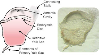
Due to more readily available samples, studies so far have focused on the first week after conception and at later stages beyond a month into pregnancy, during which organs form and mature. However, there is currently very little understanding of events that take place in the intervening days, which includes the crucial gastrulation stage that occurs shortly after the embryo implants in the womb.

Analysis of a unique sample by researchers from the Department of Physiology, Anatomy and Genetics, University of Oxford and Helmholtz Zentrum München helps fill this gap in our knowledge of early human embryogenesis. Their findings, published in the journal Nature, will contribute to the improvement of experimental stem cell models.

Gastrulation is one of the most critical steps of development, and takes place roughly between days 14 and 21 after fertilization. A single-layered embryo is transformed into a multi-layered structure known as the gastrula. During this stage, the three main cell layers that will later give rise to the human body’s tissues, organs and systems are formed. Principal Investigator Professor Shankar Srinivas said: 'Our body is made up of hundreds of types of cells. It is at this stage that the foundation is laid for generating the huge variety of cells in our body – it’s like an explosion of diversity of cell types.'

Feral Hog Invasions Leave Coastal Marshes More Susceptible to Climate Change

Coastal marshes that have been invaded by feral hogs recover from disturbances up to three times slower than non-invaded marshes and are far less resilient to sea-level rise, extreme drought and other impacts of climate change, a new study led by scientists at Duke University and the University of Massachusetts Boston (UMB) finds.

“Under normal circumstances, marshes can handle and recover from drought or sea level rise, given time, but there is no safety net in place for hog invasions,” said Brian Silliman, Rachel Carson Distinguished Professor of Marine Conservation Biology at Duke, who co-authored the study.

“Marshes that are invaded by hogs recover slower from drought, are less resilient to erosion, and hemorrhage carbon dioxide back into the air as hogs turn vast areas of the marsh into mud pits,” Silliman said.

“Based on data from our experiments, our disturbance-recovery model suggests full marsh recovery could take an extra 80 to 100 years,” he said.

Feral hogs are ravenous predators with an insatiable hunger for ribbed mussels, a shellfish species that is one of the most common – and ecologically important – inhabitants of southeastern salt marshes.

There May Be More Bird Species in The Tropics Than We Know

White-crowned Manakin
Image Credit: Phillip Edwards, Macaulay Library, Cornell Lab of ornithology. 
Study of a perky little bird suggests there may be far more avian species in the tropics than those identified so far. After a genetic study of the White-crowned Manakin, scientists say it's not just one species and one of the main drivers of its diversity is the South American landscape and its history of change. These results are published in the journal Molecular Phylogenetics and Evolution. "We found that the White-crowned Manakin probably originated in the highland forests of the Andes Mountains in northern Peru," explains lead author Jacob Berv. "Today, this bird is also found across the Amazon Basin, in the lowland rainforests of Brazil, Peru, and many other countries, including parts of Central America." Berv conducted this research while a Ph.D. student at the Cornell Lab of Ornithology and is currently a Life Sciences Fellow at the University of Michigan. "This study shows that there is a lot of evolutionary history embedded in what is commonly referred to as a 'single widespread' species in Amazonia," says co-author Camila Ribas at Brazil's National Institute of Amazonian Research. "The White-crowned Manakin is an example of a phenomenon that is probably more the rule than the exception in Amazonia—diversity is vastly underestimated by the current taxonomy."

Food waste at East Coast Lagoon Food Village to be turned into energy and fertilizer under pilot project

Associate Professor Tong Yen Wah, who leads the NUS team,
is pictured next to the anaerobic digester at the
East Coast Lagoon Food Village.
An anaerobic digestion system for food waste treatment is being piloted at the East Coast Lagoon Food Village. The system was developed by a team of researchers from the National University of Singapore (NUS) and converts food waste generated by food stalls and patrons at East Coast Lagoon Food Village into biogas and bio-fertilizer. A biogas engine converts the biogas into electricity, while the bio-fertilizer is used in landscaping applications. The onsite treatment of food waste reduces the need to send food waste for incineration.

Food waste is one of the priority waste streams identified under Singapore’s Zero Waste Masterplan. In 2020, food waste accounted for about 11 per cent of the total waste generated in Singapore, but only 19 per cent of the food waste was recycled. The rest of it was disposed of at waste-to-energy (WTE) plants. Therefore, reducing food wastage, redistributing unsold or excess food, and recycling/treating food waste are important food waste management strategies. Food waste needs to be managed holistically, as it can contaminate other recyclables when they are disposed of together, making the recyclables unsuitable or difficult to recycle. It can also give rise to odor nuisance and vermin proliferation issues, if not managed properly or in a timely manner.

As part of efforts to treat food waste and demonstrate the feasibility of on-site food waste treatment, the National Environment Agency (NEA) is supporting NUS in conducting a pilot trial of their containerized Anaerobic Digestion system at East Coast Lagoon Food Village, under the Closing the Waste Loop (CTWL) R&D Initiative. The NUS team, led by Associate Professor Tong Yen Wah from the NUS Department of Chemical and Biomolecular Engineering, oversees the operation and maintenance of the Anaerobic Digestion system. The team is concurrently studying the human psychology and behavioral factors in encouraging hawkers and cleaners to segregate food waste from other waste.

Fraternizing vampire bats share 'social microbiomes'

Microbes that make their homes on the skin or in the digestive tracts of animals can be beneficial or pathogenic to the individual and to the community. A new study finds that the gut microbiomes of vampire bats become more similar the more often the bats engage in social behaviors with one another.  Image Credit: Uwe Schmidt
In an unusual study, researchers brought vampire bats from distant Panamanian populations together for four months in a laboratory setting and tracked how the bats’ gut microbes changed over time. They found that bats that interacted closely with one another shared much more than body heat.

Reported in the journal Biology Letters, the study revealed that the gut microbiomes of bats became more similar the more often they engaged in social behaviors with one another. Such behaviors included huddling together for warmth, grooming themselves and their neighbors, and – in rare cases – sharing food via regurgitation.


This is the first study of social microbiomes to control for other factors – such as diet and environment – that could contribute to microbiome similarities, the researchers said. The study kept all the bats together in one enclosure and the bats consumed the same, laboratory-prepared food: cattle and pig blood.

Tuesday, November 16, 2021

Research Reveals How to Design a Better Next-Generation Lithium-Ion Battery

A solid-state lithium-ion battery is composed of an anode, a cathode, and a solid electrolyte separating the two. Rapidly cycling (repeatedly charging and discharging) a lithium-ion battery limits the battery's performance over time by significantly increasing the battery's internal impedance (its time-dependent resistance), which hinders the flow of current. NIST researchers, in collaboration with Sandia National Laboratories, have combined two complementary techniques – contact potential difference measurements and neutron depth profiling – to precisely determine which parts of the battery contribute most to its impedance. 
Credit: S. Kelley/NIST

The newest generation of lithium-ion batteries now under development promises a revolution in powering cell phones, electric vehicles, laptops and myriad other devices. Featuring all solid-state, nonflammable components, the new batteries are lighter, hold their charge longer, recharge faster and are safer to use than conventional lithium-ion batteries, which contain a gel that can catch on fire.

However, like all batteries, solid-state lithium-ion batteries have a drawback: Due to electro-chemical interactions, impedance--the AC analog of DC electrical resistance--can build up within the batteries, limiting the flow of electric current. Researchers at the National Institute of Standards and Technology (NIST) and their colleagues have now pinpointed the location where most of this buildup occurs. In so doing, the team has suggested a simple redesign that could dramatically limit the buildup of impedance, enabling the batteries to fulfill their role as the next-generation power source.

A better-fitting molecular ‘belt’ for making new drugs

David Nagib
The most common pharmaceuticals on the market are made by chaining together rings of molecules to create the drugs that treat conditions including pain, depression and leukemia.

But creating those rings and forming them in a way that is tailored to each individual disease has always been a cumbersome and expensive process in medicinal chemistry.

New research, published today in the journal Chem, proposes a way to simplify that transformation. The discovery will likely make it easier to produce new drug candidates, the researchers say.

David Nagib, senior author of the study and associate professor of chemistry at The Ohio State University, likened the chain of molecules to a belt with no holes: With no way to fasten the circle and no measurements for where holes might go, the belt can’t be assembled in a way that keeps it closed.

“The problem we were trying to solve is how do you punch the hole so that it fits you perfectly, and get it right on the first try without measuring,” Nagib said. “The trick here was we had to put the holes in just the right place, but we had to figure out precisely where the holes should go, without any markings to tell us where that might be.”

The “belt” in this case is a string of carbon-hydrogen bonds, the most ubiquitous bonds in all of nature and medicines. Most drugs contain rings of carbon-hydrogen bonds, linked together by a “bridging” nitrogen atom, within complex structures that interact precisely with cellular components in the body – like a key fitting into a lock. The most common ring found in all medications are six-sided ones, called a piperidine.

But piperidines have long been difficult and expensive to produce, primarily because chemists could not quickly or cheaply replace a carbon-hydrogen bond with other chemical bonds.

Human Proteoform Project to map proteins in human body


Project will accelerate the development of precision diagnostics and therapeutics

Now that the Human Genome Project has officially wrapped, an international team of researchers will map the entire collection of proteins in the human body.

Plans and goals for the Human Proteoform Project were outlined in a paper published last in the journal Science Advances. The large undertaking will characterize known proteoforms (specific protein molecules) as well as aim to systematically discover and analyze new ones in human tissues, cells and fluids.

“We are all built of proteins, and most drugs target proteins,” said Northwestern University’s Neil Kelleher, a world-renowned proteomics pioneer and corresponding author of the paper. “But understanding proteins is an open frontier. Like other seminal moments in science and technology, this project will serve as a major achievement that can help us more fully understand proteins’ role in all types of disease, aging and new therapeutics.”

Sandia cooks material-storage containers to assess fire safety

A test where a stainless-steel container designed for the storage and transportation of hazardous materials is heated to 2000 degrees Fahrenheit for four hours at Sandia National Laboratories. The container did not catastrophically fail, instead small pinholes formed relieving the intense pressure. On the upper left-hand corner is an X-ray video from outside the container. The top middle image is a video from a pinhole camera inside the test chamber. The upper right-hand corner shows the outside of the test chamber. The bottom graph shows the pressure inside the container (white line) and the temperature of the container (in degrees Celsius) from nine different places (colored lines) during the test. 
Video Credit: Sandia National Laboratories

A team at Sandia National Laboratories has completed a series of tests on specially designed stainless-steel containers used by the Department of Energy for storage and transportation of hazardous materials.

The engineers, technologists and project managers were surprised to find that the containers did not split open when heated to 2000 degrees Fahrenheit. That is almost as hot as a cement kiln.

“These containers were welded shut and heated to 2000 degrees, so we assumed that they were going to split open, but they developed small pinholes instead,” said Walt Gill, the test director and Sandia mechanical engineer. “We think the material inside reacted with the container itself and produced the pinholes in the container. These tiny holes let out all of the superheated gas without the containers pressurizing and pulling themselves apart.”

The series of 10 tests were designed to mimic a hypothetical raging-hot fire burning at a DOE facility and engulfing a container that had been knocked on its side and left outside of its insulated packaging, which protects it from heat. Since these containers are not designed to withstand such a fire, the goal of the test was to determine how much, if any, material stored within the container would be released into the air during such an accident, said Gill and Austin Baird, the test engineer.

Map of Mouse Iris Offers New Look at the Eye

Branching nerve fibers (red) stretch across the mouse iris. Scientists have now built the first cell-by-cell map of this eye tissue.
Credit: J. Wang

A cell-by-cell map of the mouse iris lays the foundation to study eye disorders and engineer cell therapies to replace damaged eye tissue.

If vision science were a movie, the iris would be a supporting actor. It doesn’t get as much limelight as the retina, the eye’s light-sensing tissue. And it’s not as high-profile as the lens, which can cloud with cataracts as people age.

Though the iris – the colorful tissue that rings the pupil – comes in a rainbow of showy shades, for most scientists, “it was not the main attraction in the eye,” says Jeremy Nathans, a Howard Hughes Medical Institute Investigator at Johns Hopkins University School of Medicine. In fact, he has spent most of his career studying the molecular biology of the retina.

“The basic biology of the iris had kind of languished,” Nathans says. Not anymore. He and his colleagues Jie Wang and Amir Rattner have now developed a high-resolution map of the mouse iris that distinguishes individual cells by the activity of their genes. The trio was motivated by the beauty of the iris, the diversity of iris structure in different animals, and its importance for vision, Nathans says.

Strands of dilator muscles (red) weave through the mouse iris.
Sphincter cells (green) cluster in the pupil (right),
which expands and constricts to control the amount of light entering the eye.
Credit: J. Wang
His team identified eight types of iris cells and uncovered new information about iris cell development and the genes that switch on when the pupil dilates, they report November 16, 2021, in the journal eLife. The researchers’ cell-by-cell map of the iris could one day help identify genes involved in iris-related eye disorders. The work could also guide engineering of healthy iris cells used to replace diseased cells elsewhere in the eye.

Featured Article

Discovery of unexpected collagen structure could ‘reshape biomedical research’

Jeffrey Hartgerink is a professor of chemistry and bioengineering at Rice. Photo Credit: Courtesy of Jeffrey Hartgerink / Rice University Co...

Top Viewed Articles