 |
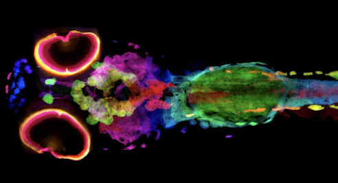
| 3D image of melanin in a zebrafish sample captured by micro-computed tomography. Credit: Spencer R. Katz and Daniel J. Vanselow/Penn State College of Medicine |
Researchers have developed a new technique that images every pigment cell of a whole zebrafish in 3D. The work, recently reported in the journal eLife, could help scientists understand the role of melanin in skin cancer.
Melanin is a natural pigment that gives color to the skin, hair, and eyes in humans and animals. Melanin also has implications in melanin-containing cancers, or melanomas, which are typically staged by the depth of penetration in skin.
But studying melanin directly with a conventional microscope is challenging because the pigment blocks light. So Keith C. Cheng, a distinguished professor of pathology, pharmacology and biochemistry, and molecular biology at Penn State College of Medicine, turned to X-ray imaging, which can pass through optically opaque matter like melanin.
To perform the imaging, Cheng partnered with Dula Parkinson, a staff scientist at Berkeley Lab’s Advanced Light Source (ALS), to image two sets of zebrafish samples – one with the normal pigmentation associated with the zebrafish’s characteristic black stripes, and another from a mutant zebrafish line with lighter stripes called golden. Over 15 years ago, Cheng and his lab discovered a key gene implicated in human skin color by studying golden zebrafish. That discovery highlighted the zebrafish’s utility as an animal model of human pigmentation in skin disorders such as albinism or melanoma skin cancer.


.jpg)