. Scientific Frontline

Thursday, June 9, 2022

Europe’s Largest Land Predator Unearthed

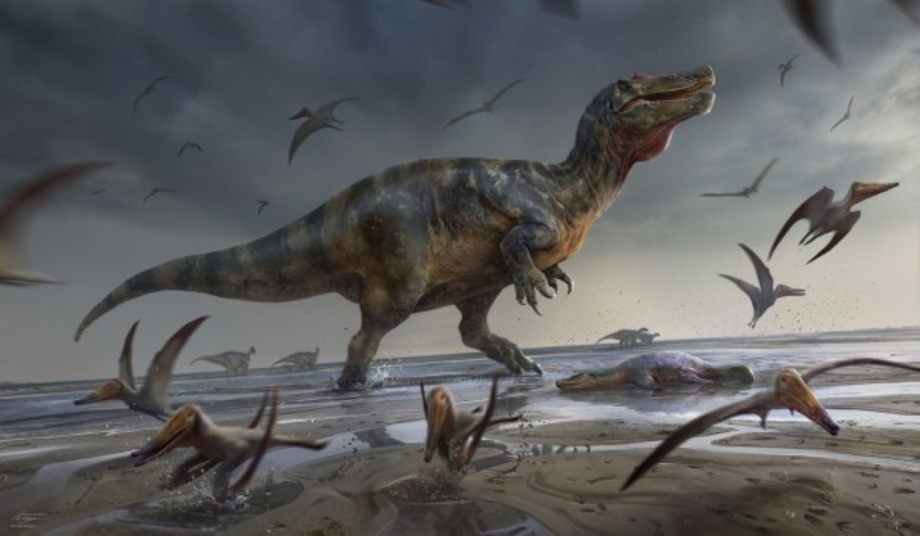
Illustration of White Rock spinosaurid.
Credit: Anthony Hutchings 

Research involving paleontologists from the Universities of Portsmouth and Southampton has identified the remains of one of Europe’s largest ever land-based hunters: a dinosaur that measured over 10m long and lived around 125 million years ago.

Several prehistoric bones, uncovered on the Isle of Wight, on the south coast of England, and housed at Dinosaur Isle Museum in Sandown, belonged to a type of two-legged, crocodile-faced predatory dinosaur known as spinosaurids. Dubbed the ‘White Rock spinosaurid’ – after the geological layer in which it was found – it was a predator of impressive proportions.

“This was a huge animal, exceeding 10 m in length and probably several tons in weight. Judging from some of the dimensions, it appears to represent one of the largest predatory dinosaurs ever found in Europe – maybe even the biggest yet known”, said University of Southampton PhD student Chris Barker, who led the study. “It’s a shame it’s only known from a small amount of material, but these are enough to show it was an immense creature.”

The discovery follows previous work on spinosaurids by the University of Southampton team, which published a study on the discovery of two new species in 2021.

Social isolation may impact brain volume in regions linked to higher risk of dementia

Elderly woman in the middle stages of Alzheimer 
Credit: Steven HWG

Social isolation is linked to lower brain volume in areas related to cognition and a higher risk of dementia, according to research published today in Neurology. The study found that social isolation was linked to a 26% increased risk of dementia, separately from risk factors like depression and loneliness.

“Social isolation is a serious yet underrecognized public health problem that is often associated with old age,” said study author Professor Jianfeng Feng of Fudan University in Shanghai, China. “In the context of the COVID-19 pandemic, social isolation, or the state of being cut off from social networks, has intensified. It’s more important than ever to identify people who are socially isolated and provide resources to help them make connections in their community.”

The study looked at over 460,000 people across the United Kingdom with an average age of 57 at the beginning of the study who were followed for nearly 12 years before the pandemic. Of those, almost 42,000 (9%) reported being socially isolated, and 29,000 (6%) felt lonely. During the study, almost 5,000 developed dementia.

Researchers collected survey data from participants, along with a variety of physical and biological measurements, including MRI data. Participants also took thinking and memory tests to assess their cognitive function. For social isolation, people were asked three questions about social contact: whether they lived with others; whether they had visits with friends or family at least once a month; and whether they participated in social activities such as clubs, meetings or volunteer work at least once a week. People were considered socially isolated if they answered no to at least two questions.

Visual system brain development implicated in infants who develop autism

Anatomical locations of the splenium (yellow) and right middle occipital gyrus (red) in a representative infant brain.
Source: University of North Carolina at Chapel Hill

For the first time, scientists have found that brain differences in the visual brain systems of infants who later develop autism are associated with inherited genetic factors.

Published in the American Journal of Psychiatry, this research shows that brain changes in the size, white matter integrity and functional connectivity of the visual processing systems of six-month-olds are evident well before they show symptoms of autism as toddlers. Moreover, the presence of brain changes in the visual system is associated with the severity of autism traits in their older siblings.

Led by Dr. Jessica Girault, assistant professor of psychiatry at the UNC School of Medicine, this is the first research to observe that infants with older siblings who have autism and who themselves later develop autism as toddlers have specific biological differences in visual processing regions of the brain and that these brain characteristics precede the appearance of autistic symptoms. The presence of those visual processing differences is related to how pronounced the autism traits are in the older siblings.

“We’re beginning to parse differences in infant brain development that might be related to genetic factors,” said Girault, who is also a member of the Carolina Institute of Developmental Disabilities. “Using magnetic resonance imaging, we studied selected structures of brain, the functional relationship between key brain regions, and the microstructure of white matter connections between those brain regions. Findings from all three pointed us to the discovery of unique differences in the visual systems of infants who later developed autism.”

Higher rate of COVID-19 death before vaccination linked to certain common inflammatory immune conditions

People with certain inflammatory immune conditions affecting the joints, bowel and skin, such as rheumatoid arthritis, may have been more at risk of dying or needing hospital care if they got COVID-19 before vaccination compared with the general population, according to a new study published in The Lancet Rheumatology with the involvement of researchers from the University of Oxford.

The findings are based on an analysis of 17 million patient GP records in England during the first phase of the pandemic from March-September 2020, when the UK was in lockdown and before vaccines were available. Since then, many of the people treated with medicines analyzed in this study have been specifically targeted for third primary vaccine doses followed by boosters and are on a list of people to offered anti-viral treatments.

The study was conducted by a team from the London School of Hygiene & Tropical Medicine (LSHTM) and the University of Oxford using the OpenSAFELY platform along with colleagues from St John’s Institute of Dermatology at Guy’s and St Thomas’ NHS Foundation Trust, King’s College London, the University of Exeter and University of Edinburgh.

More than 1 million patients in the analysis had immune mediated inflammatory diseases (IMIDs). These included inflammatory bowel disease such as Crohn’s disease and ulcerative colitis, conditions affecting the joints such as rheumatoid arthritis, and skin conditions including psoriasis.

Invasive Insect that Kills Grapes Could Reach California Wine Region by 2027

The invasive insect, the spotted lanternfly.
Credit: U.S. Department of Agriculture.

The spotted lanternfly, an invasive insect that can kill grapevines and damage other crops, has a chance of first reaching the wine-producing counties of California in five years, according to a new analysis from North Carolina State University researchers.

In the study published in Communications Biology, researchers used a computer simulation tool to predict the timing of the spread of the spotted lanternfly, Lycorma delicatula, across the United States if efforts to control its spread are stopped. They predicted there is a high probability of the insect spreading to North Carolina by 2027, and a chance of the insect first reaching California’s grape-producing counties that same year.

“This is a big concern for grape growers; it could lead to billions of dollars of losses in the agricultural sector,” said the study’s lead author Chris Jones, research scholar with the NC State Center for Geospatial Analytics. “With this study, we have a baseline that we can use to evaluate the effect of different management strategies.”

The spotted lanternfly is native to Asia. It was first identified in the United States in Pennsylvania in 2014. Since then, it has spread to at least 11 other states. The invasive insect can damage or destroy commercially valuable crops such as grapes, apples, almonds, walnuts, cherries, hops, and peaches, as well as certain trees. It kills plants by directly feeding on them, and can also damage them by leaving behind a residue known as “honeydew” that helps mold grow. California, which produces 82% of the nation’s grapes, has been identified along with Washington state as a “highly suitable” climate for the spotted lanternfly.

Multi-scale imaging confirms protein’s role in neuronal structure, dynamics

A whole, live cell time-lapse image (2.5 min) of a neuron expressing fluorescently tagged actin (green) and cofilin (red).
Credit: Penn State College of Medicine.

Protein structures are typically determined by studying them in their purified form, outside of the busy inner workings of the cell, and because of this, their biological relevance is often called into question. In a new study by Penn State College of Medicine researchers, the long-observed protein structure cofilactin, a form of the filamentous protein actin that contains numerous connections to cofilin proteins, was shown to be a major component of neuronal growth cone filopodia — small dynamic “antennae” at the tips of growing neurons.

“The effects of cofilin on actin structure and its physical properties have been studied for more than 20 years, but now we can confidently see that this structural alteration serves some biological function in the cell,” said Matt Swulius, assistant professor of biochemistry and molecular biology. “We’re still trying to determine the mechanistic details of its function, but we have strong evidence that cofilactin regulates the flexibility of searching filopodia.”

According to Swulius, understanding how filopodial dynamics are controlled at the molecular level could open therapeutic avenues into nerve regeneration as well as some developmental diseases. His lab is studying how the proteins fascin and cofilin function together to regulate the structure and movement of neuronal filopodia as they navigate their environment to eventually form cellular connections.

New Light on Magnetic Massive Stars

Video Credit: Jeffrey C. Chase

Oh, those twinkling, twinkling little stars. They make the night sky seem so peaceful, so idyllic, so quiet. It’s hard to believe that they are actually powerful furnaces, where explosive power, extreme heat and swirling, energized gases make them something more like a hydrogen bomb than anything imagined in that child’s lullaby.

Some of us settle for the wistful bit of that lullaby: “How I wonder what you are.” Others press on with increasingly challenging questions in a lifelong quest to learn, discover and inform the rest of us about what those twinkling little stars really are.

Matt Shultz does that as the Annie Jump Cannon Postdoctoral Fellow at the University of Delaware. Stan Owocki does that as professor of physics and astronomy at UD. With collaborators at UD and around the world, they recently joined forces on three articles printed by the Royal Astronomical Society, pointing to important new discoveries that could change how measurements of stars are done and how their brightness can be predicted.

It started a few years ago. Shultz was minding his astronomy at UD and Paolo Leto, a researcher at the Catania Astrophysical Observatory, was doing the same in Italy. Both were studying magnetic massive stars — a rare variety of bright, hot stars with magnetic fields thousands of times stronger than the sun’s. These stars are emerging as promising laboratories for studying plasmas under extreme conditions, Shultz said.

Ground-breaking number of brown dwarfs discovered

Image of the brown dwarf (in the red circle) discovered around the star HIP 21152, obtained with the Very Large Telescope SPHERE instrument.
Credit: M. Bonavita et al., MNRAS

Brown dwarfs, mysterious objects that straddle the line between stars and planets, are essential to our understanding of both stellar and planetary populations. However, only 40 brown dwarfs could be imaged around stars in almost three decades of searches. An international team led by researchers from the Open University and the University of Bern directly imaged a remarkable four new brown dwarfs thanks to a new innovative search method.

Brown dwarfs are mysterious astronomical objects that fill the gap between the heaviest planets and the lightest stars, with a mix of stellar and planetary characteristics. Due to this hybrid nature, these puzzling objects are crucial to improve our understanding of both stars and giant planets. Brown dwarfs orbiting a parent star from sufficiently far away are particularly valuable as they can be directly photographed – unlike those that are too close to their star and are thus hidden by its brightness. This provides scientists with a unique opportunity to study the details of the cold, planet-like atmospheres of brown dwarf companions.

Wednesday, June 8, 2022

Evasive quantum phenomenon makes debut in routine tabletop experiment

Researchers recently confirmed the presence of the axial Higgs mode, a particle excitation depicted here as a golden sphere. They used Raman spectroscopy, in which an incoming electric field, shown in blue, was coupled with the particle and subsequently scattered into a different frequency, shown in red.
 Credit: Ioannis Petrides and Prineha Narang/Harvard University

A Quantum Science Center-supported team has captured the first-ever appearance of a previously undetectable quantum excitation known as the axial Higgs mode.

This mode manifests as a low-energy excitation in rare-earth tellurides, a class of quantum materials notable for exhibiting charge density wave, or CDW, interactions. This behavior refers to arrangements of interacting electrons in quantum materials that form specific patterns and correlations.

Unlike the regular Higgs mode, which is produced by a Higgs mechanism that provides mass to fundamental particles in the Standard Model of Particle Physics, the axial Higgs mode is visible at room temperature. This characteristic enables more efficient and cost-effective experiments for manipulating quantum materials for various applications – including next-generation memory storage and opto-electronic devices – which would otherwise require extremely cold temperatures.

The team responsible for these results, which are published in Nature, was led by researchers at Boston College and includes scientists from Harvard University, Princeton University, University of Massachusetts Amherst, Yale University, University of Washington and the Chinese Academy of Sciences.

New delivery method allows slow-release of broader array of peptide drugs in the body

Schwendeman Lab.
Image credit: Michigan Photography

A new study from the University of Michigan describes one of the first entirely new drug delivery microencapsulation approaches in decades.

Microencapsulation in biodegradable polymers allows drugs such as peptide therapeutics to be released over time in the body.

Peptides are molecules in the body that are composed of short chains of amino acids, and include messengers, growth factors and well-known hormones such as insulin. Because of their larger size and structure, peptide drugs are rarely given by mouth and must be injected. Microencapsulation is one way to decrease the time needed between injections.

One slow-release delivery method for peptide drugs is to encapsulate them within the type of resorbable polymers often used as dissolving sutures, said study co-author Steven Schwendeman, professor of pharmaceutical sciences and biomedical engineering.

However, development of polymer dosage forms for delivery of certain peptide drugs has been difficult because the currently available methods to microencapsulate the peptide molecules in the polymer require organic solvents and complex manufacturing.

Featured Article

Discovery of unexpected collagen structure could ‘reshape biomedical research’

Jeffrey Hartgerink is a professor of chemistry and bioengineering at Rice. Photo Credit: Courtesy of Jeffrey Hartgerink / Rice University Co...

Top Viewed Articles