. Scientific Frontline

Thursday, July 28, 2022

Carbon Removal Using ‘Blue Carbon’ Habitats “Uncertain and Unreliable”


Restoring coastal vegetation – so called ‘blue carbon’ habitats – may not be the nature-based climate solution it is claimed to be, according to a new study.

In their analysis researchers from the University of East Anglia (UEA), the French Centre National de la Recherche Scientifique (CNRS) and the OACIS initiative of the Prince Albert II of Monaco Foundation, challenge the widely held view that restoring areas such as mangroves, saltmarsh and seagrass can remove large amounts of carbon dioxide (CO2) from the atmosphere.

The findings of their review, published today in the journal Frontiers in Climate, identify seven reasons why carbon accounting for coastal ecosystems is not only extremely challenging but risky.

These include the high variability in carbon burial rates, vulnerability to future climate change, and fluxes of methane and nitrous oxide. The authors, who also looked at information on restoration costs, warn that extra measurements can reduce these risks, but would mean much higher costs.

However, they stress that blue carbon habitats should still be protected and, where possible, restored, as they have benefits for climate adaptation, coastal protection, food provision and biodiversity conservation.

Lead author Dr Phil Williamson, honorary reader in UEA’s School of Environmental Sciences, said: “We have looked into the processes involved in carbon removal and there are just too many uncertainties. The expected climate benefits from blue carbon ecosystem restoration may be achieved, yet it seems more likely they will fall seriously short.

New rabies vaccine candidate demonstrates promising immune response and safety

Artist's impression of the rabies virus
Source: University of Oxford

Researchers from the University of Oxford have today reported new findings from a Phase 1 clinical trial studying the immune response and safety of their newly-developed single shot rabies vaccine, ChAdOx2 RabG - with promising results identified.

The RAB001 trial was conducted at the University and is the first time the novel rabies vaccine has been used in human volunteers. The aim of the study was to look at safety and measure immune responses from the vaccine by analyzing levels of rabies neutralizing antibodies – a powerful marker of successful rabies vaccination.

In their findings (published in The Lancet Microbe), the researchers reported that 12 volunteers were recruited into the study in total, with three receiving a low dose, three receiving a medium dose and six receiving a high dose of ChAdOx2 RabG. Strong immune responses against rabies were generated by the vaccine, with all volunteers who received a medium or high dose developing levels of rabies neutralizing antibodies above the World Health Organization protective threshold (0.5 International Units / ml) within two months.

No serious adverse events or safety concerns were reported during the trial. Expected levels of common short-lived vaccine side effects such as soreness at the injection area or feverishness were observed in volunteers, mainly in the medium- and higher-dose groups.

Additionally, the researchers assessed longer term immune responses. Six of the seven middle- and high-dose recipients who returned for an additional follow-up one year after vaccination maintained neutralizing antibody levels above the protective threshold, demonstrating that the immune response from the vaccine persists over time.

Coming wave of opioid overdoses 'will be worse than ever been before'

Source/Credit: Northwestern University

Over the past 21 years of opioid overdose deaths—from prescription drugs to heroin to synthetic and semisynthetic opioids such as fentanyl—geography has played a role in where opioid-involved overdose deaths have occurred, reports a new Northwestern Medicine study.

"For the first time, there is a convergence and escalation of acceleration rates for every type of rural and urban county.”
Lori Post
Director of the Buehler Center for Health Policy and Economics, Northwestern University Feinberg School of Medicine

But the coming wave will not discriminate between rural and urban areas, the study findings suggest. Every type of county—from the most rural to the most urban—is predicted to see dramatic increases in deaths from opioid-involved overdoses. The reason opioid overdoses have reached historical highs comes from combining synthetic opioids with stimulants such as cocaine and methamphetamines, a lethal cocktail that is hard to reverse during an overdose, the study authors said.

“I'm sounding the alarm because, for the first time, there is a convergence and escalation of acceleration rates for every type of rural and urban county,” said corresponding author Lori Post, director of the Buehler Center for Health Policy and Economics at Northwestern University Feinberg School of Medicine. “Not only is the death rate from an opioid at an all-time high, but the acceleration of that death rate signals explosive exponential growth that is even larger than an already historic high.”

MIT engineers develop stickers that can see inside the body


Ultrasound imaging is a safe and noninvasive window into the body’s workings, providing clinicians with live images of a patient’s internal organs. To capture these images, trained technicians manipulate ultrasound wands and probes to direct sound waves into the body. These waves reflect back out to produce high-resolution images of a patient’s heart, lungs, and other deep organs.

Currently, ultrasound imaging requires bulky and specialized equipment available only in hospitals and doctor’s offices. But a new design by MIT engineers might make the technology as wearable and accessible as buying Band-Aids at the pharmacy.

In a paper appearing today in Science, the engineers present the design for a new ultrasound sticker — a stamp-sized device that sticks to skin and can provide continuous ultrasound imaging of internal organs for 48 hours.

The researchers applied the stickers to volunteers and showed the devices produced live, high-resolution images of major blood vessels and deeper organs such as the heart, lungs, and stomach. The stickers maintained a strong adhesion and captured changes in underlying organs as volunteers performed various activities, including sitting, standing, jogging, and biking.

The current design requires connecting the stickers to instruments that translate the reflected sound waves into images. The researchers point out that even in their current form, the stickers could have immediate applications: For instance, the devices could be applied to patients in the hospital, similar to heart-monitoring EKG stickers, and could continuously image internal organs without requiring a technician to hold a probe in place for long periods of time.

Boeing, U.S. Air Force Celebrate 50 Years of F-15 Innovation

F-15 Tower flyby
Credit: Boeing

On July 27, 1972, the Boeing [NYSE: BA] F-15 flew for the first time with Chief Test Pilot Irv Burrows at the controls. Fifty years later, the undefeated F-15 continues to evolve and add advanced capability to the U.S. Air Force fighter fleet.

“Boeing is proud of the F-15’s proven performance and of our shared legacy on this platform with the U.S. Air Force and operators around the world,” said Prat Kumar, vice president of F-15 Programs. “With its unrivaled combat performance, five decades-long production run and continuous evolution, the F-15 has a remarkable history and continues today to be a critical asset for U.S. and allied forces. And with the development of new, advanced capabilities and the evolution of the F-15EX, the best is yet to come.”

Boeing’s F-15 program was initiated at the request of the U.S. Air Force, which needed a fighter jet designed to maintain the country’s air superiority. Through its variants, the F-15 has also served that mission internationally with numerous global customers including Japan, Israel, Saudi Arabia, Singapore, South Korea and Qatar.

Creating an “Adult-like” Mature Human Cardiac Tissue

Heart muscle structure, computer illustration. Heart muscle is composed of spindle-shaped cells grouped in irregular bundles. Boundaries between individual cells are faintly visible here. Each cell contains one nucleus, visible as a dark stained spot. Cardiac muscle is a specialized muscle tissue that can contract regularly and continuously without tiring.

Researchers in the Biomedical Engineering Department at UConn have developed a new cardiac cell-derived platform that closely mimics the human heart, unlocking potential for more thorough preclinical drug development and testing, and model for cardiac diseases.

The research, published in Cell Reports by Assistant Professor Kshitiz in collaboration with Dr. Junaid Afzal in the cardiology department at the University of California San Francisco, presents a method that accelerates maturation of human cardiac cells towards a state suitable enough to be a surrogate for preclinical drug testing.

“There is a very strong need to create human cardiac constructs for all sorts of applications. Small animal models just do not recapitulate human heart biology, and human samples are scarce,” says Kshitiz. “This matters because all drugs need to be tested for their toxicity to heart. It is widely believed that a large number of them unnecessarily fail clinical trials because we do not have human samples to test them with.”

Kshitiz and Afzal first identified the need to create a mature human cardiac tissue during their time together at Johns Hopkins Medicine.

Monash microbiologist to convert greenhouse gases into sustainable pet food

(L-R) Dr Rachael Lappan and A/Prof Chris Greening
Credit: Monash University

The Australian Research Council (ARC) has announced today that Monash University will receive $5 million funding to lead a new Research Hub to develop cutting-edge technologies to transform greenhouse gas emissions from the energy and manufacturing sectors into valuable products.

Monash University will partner with seven national and international academic organizations, as well as 22 industry partners including Woodside Energy, to form the ARC Research Hub for Carbon Utilization and Recycling.

Monash will use new electrochemical, thermochemical, and biochemical methods to convert the climate-active gases carbon dioxide and methane into useful products. It will also drive new policy mechanisms to support early-stage market development of products and technologies to help drive industry transformation.

The Monash arm of the biochemical conversion node will be led by Associate Professor Chris Greening, an award-winning microbiologist who heads Monash BDI’s Health in a Changing World Program.

His team will convert gases produced by the energy, agriculture, and waste sectors into protein-rich pet and fish foods. To do so, they will use bacteria that grow on gases such as methane, carbon dioxide, and hydrogen alone.

AI tackles the challenge of materials structure prediction


The researchers from Cambridge and Linkoping Universities, have designed a way to predict the structure of materials given its constitutive elements. The results are reported in the journal Science Advances.

The arrangement of atoms in a material determines its properties. The ability to predict this arrangement computationally for different combinations of elements, without having to make the material in the lab, would enable researchers to quickly design and improve materials. This paves the way for advances such as better batteries and photovoltaics.

However, there are many ways that atoms can ‘pack’ into a material: some packings are stable, others are not. Determining the stability of a packing is computationally intensive, and calculating every possible arrangement of atoms to find the best one is not practical. This is a significant bottleneck in materials science.

“This materials structure prediction challenge is similar to the protein folding problem in biology,” said Dr Alpha Lee from Cambridge’s Cavendish Laboratory, who co-led the research. “There are many possible structures that a material can ‘fold’ into. Except the materials science problem is perhaps even more challenging than biology because it considers a much broader set of elements.”

A Nanokelvin Microwave Freezer for Molecules

A close view inside the main vacuum chamber of the NaK molecules experiment. In the middle four high-voltage copper wires are routed to an ultrahigh-vacuum glasscell where the ultracold polar molecules were produced.
Credit: Max Planck Institute of Quantum Optics

Researchers at the Max Planck Institute of Quantum Optics have developed a novel cooling technique for molecular gases. It makes it possible to cool polar molecules down to a few nanokelvin. The trick used by the team in Garching to overcome this hurdle is based on a rotating microwave field. It helps to stabilize the collisions between the molecules during cooling by means of an energetic shield. In this way, the Max Planck researchers succeeded in cooling a gas of sodium-potassium molecules to 21 billionths of a degree above absolute zero. In doing so, they set a new low-temperature record. In the future, the new technique will allow us to create and explore many forms of quantum matter that have not been experimentally accessible until now.

When a highly diluted gas is cooled to extremely low temperatures, bizarre properties are revealed. Thus, some gases form a so-called Bose-Einstein condensate - a type of matter in which all atoms move in unison. Another example is supersolidity: a state in which matter behaves like a frictionless fluid with a periodic structure. Physicists expect to find particularly diverse and revealing forms of quantum matter when cooling gases consisting of polar molecules. They are characterized by an uneven electrical charge distribution. Unlike free atoms, they can rotate, vibrate and attract or repel each other. However, it is difficult to cool molecular gases to ultra-low temperatures.

A team of researchers at the Max Planck Institute of Quantum Optics in Garching has now found a simple and effective way to overcome this roadblock. It is based on a rotating field of microwaves.

Ural Scientists Found Earliest Evidence of Hyenas Toxocariasis

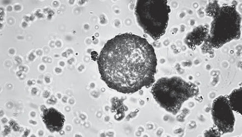
Image of a hyena coprolite taken with a microscope. In the center is a toxocara egg.
Credit: Dmitry Gimranov

Ural paleontologists, together with Permian parasitologists, found helminth eggs in coprolites (fossil excrement) of the giant short-faced hyena Pachycrocuta. This is the earliest finding indicating that this species of hyena was infected with parasites and had toxocariasis. A description of the finding and analysis of the specimens is published in Doklady Biological Sciences.

"During excavations in the Tavrida cave we found the remains of large mammals, including at least two dozen individuals of Pachycrocuta hyena, dated to the early Pleistocene (1.5-1.8 million years). We believe that hyenas used the cave Tavrida as a den for quite a long time, because here, in the southern corridor of the cave, there were a huge number of coprolites of hyenas, both single and in large assemblies. The massive teeth and especially strong enamel structure allowed hyenas to gnaw the bones of even large hoofed animals. Therefore, the Pachycrocuta could utilize the carcasses of large herbivores," says Dmitry Gimranov, Senior Researcher at the Institute of Plant and Animal Ecology of Ural Branch of Russian Academy of Sciences and Laboratory of Natural Science Methods in Humanities at Ural Federal University.

Scientists analyzed three samples of coprolites, in one of which they found parasite eggs. Based on the size and morphology, paleontologists determined that these were helminth eggs. Scientists believe that toxocariasis was a widespread disease among extinct hyenas. This is also confirmed by the data of other researchers. Eggs of helminths of 1.2 million years old were found in coprolites of the same hyena species from the Haro site in Pakistan and 0.3-0.5 million years old at the Menez-Dregan site in France. There are also finds in Italy (Costa San Gicomo site) dated at 1.5 million years. The find in Tavrida will not only help to complete the list of parasites of ancient animals and compare it with helminths of modern hyenas, but also to clarify other features of ancient animals.

"Ancient animal coprolites are unique fossils reflecting biological features that cannot be demonstrated by studying bone remains. Coprolites can be a valuable source of paleoclimate data because they may contain pollen and spore remains of ancient plants. Coprolites may also contain remains of ancient parasites, which provides a unique opportunity to obtain additional information about the ecology of extinct species," adds Daniyar Khantemirov, Laboratory of Natural Science Methods in Humanities researcher.

Note that the research team included employees of the Ural Federal University, the Institute of plant and animal ecology Ural Branch of the Russian Academy of Sciences and the Perm State Agro-Technological University named after Academician D.N. Pryanishnikov.

Reference:
Toxocariasis is an infection caused by animal ascarid larvae. Other helminth eggs of toxocarias mature in the soil and infect dogs, cats and other animals. The source of the disease, toxocara was discovered by the German scientist Werner in 1782. Only in 1950 lesion with these helminths was isolated as a separate disease. Eggs from toxocars can be found in the ground and contaminated water.

Source/Credit: Ural Federal University

pal072822_01

Featured Article

Discovery of unexpected collagen structure could ‘reshape biomedical research’

Jeffrey Hartgerink is a professor of chemistry and bioengineering at Rice. Photo Credit: Courtesy of Jeffrey Hartgerink / Rice University Co...

Top Viewed Articles