. Scientific Frontline

Monday, October 24, 2022

A revolutionary method to observe cell transport

Nanobodies (grey) with magnetic probes (red stars) target the desired membrane protein.
Credit: Bordignon, Enrica

Membrane proteins are key targets for many drugs. They are located between the outside and inside of our cells. Some of them, called ‘‘transporters’’, move certain substances in and out of the cellular environment. Yet, extracting and storing them for observation is particularly complex. A team from the University of Geneva (UNIGE), in collaboration with the University of Zurich (UZH), has developed an innovative method to study their structure in their native environment: the cell. The technique is based on electron spin resonance spectroscopy. These results, just published in the journal Science Advances, may facilitate future development of new drugs.

In living organisms, each cell is surrounded by a cell membrane (or ‘‘cytoplasmic membrane’’). This membrane consists of a double layer of lipids. It separates the contents of the cell from its direct environment and regulates the substances that can enter or leave the cell. The proteins attached to this membrane are called ‘‘membrane proteins’’.

Located at the interface between the outside and inside of the cell, they carry various substances across the membrane - into or out of the cell - and play a crucial role in cell signaling, i.e. in the communication system of cells that allows them to coordinate their metabolic processes, development and organization. As a result, membrane proteins represent more than 60% of current drug targets.

Using Carbon-Carbon Clumping to Detect the Signature of Biotic Hydrocarbons


The mystery of the origin of hydrocarbons found in extraterrestrial environment may finally be resolved, thanks to a technique developed by researchers at Tokyo Tech based on a 13C-13C abundance analysis. By measuring the abundance of clumped 13C-13C isotope in the hydrocarbons, it can be inferred if a hydrocarbon was produced via biological processes. This could open doors to distinguishing such hydrocarbons from abiotic ones, aiding our search for extra-terrestrial life.

An important signature of life is the existence of organic molecules that have originated from biological processes. The most common organic molecule found in all life forms are hydrocarbons. However, they need not be of biotic origin, i.e., produced from thermal decomposition of sedimentary organic matter or microbes. So, while hydrocarbons have been found in several places outside Earth, they are not necessarily indicative of extra-terrestrial life. These hydrocarbons could well have formed from abiotic, or non-biological processes. Therefore, finding out whether a hydrocarbon is of biotic or abiotic origin is key to inferring the existence of life. Unfortunately, this has proved to be a tremendously challenging task so far.

How heart failure disrupts the cell’s powerhouse

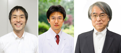
From left: Shingo Takada, Hokkaido University and Hokusho University; Shintaro Kinugawa, Kyushu University; and Hisataka Sabe, Hokkaido University
Photos credits: Shingo Takada, Shintaro Kinugawa, Hisataka Sabe

Chronic heart failure causes the cell’s powerhouses to dysfunction, in part due to overconsumption of an important intermediary compound in energy production. Supplementing the diet to compensate for this could prove a promising strategy for treating heart failure. The findings were published in the journal PNAS by Hokkaido University scientists and colleagues in Japan.

Mitochondria are small organelles found in almost every cell and are responsible for converting carbohydrates, fats and proteins into energy to power biochemical reactions. Chronic heart failure is known to be associated with mitochondrial dysfunction, but much is still unknown about how this happens at the molecular level.

A research team consisting of molecular biologist Hisataka Sabe (Hokkaido University), cardiovascular medicine specialists Shingo Takada (Hokkaido University and Hokusho University) and Shintaro Kinugawa (Kyushu University) and their colleagues studied the biochemical processes that occur in mice with chronic heart failure caused by surgically blocking part of the blood supply to their hearts. They specifically looked at heart cells outside the boundaries of dead tissue.

Saturday, October 22, 2022

Aging, Frailty, and our Microbiomes

Photo Credit: Magda Ehlers

We humans tend to think we live independently, capable of ensuring our own health and wellbeing. As researchers are increasingly aware, however, our microbiomes—the trillions of microbes that live on and within us—play central roles in our health and susceptibility to different diseases. And as we age, our microbiomes change too, with important health implications over time.

Jackson Laboratory (JAX) Associate Professor Julia Oh, Ph.D., studies the microbiome, particularly the microbes that colonize the skin. While prior research has explored the gut microbiome in the context of aging, to date there has been little insight into the changes that occur in other microbial communities of our body, like the mouth and skin. To further investigate, Oh and her team collaborated with UConn Center on Aging Professors Julie Robison, Ph.D., and George Kuchel, M.D., to study the microbiome of the skin, oral, and gut of older adults compared to younger adults.

Because of the unique design of their study, where they sampled frail older adults inhabiting skilled nursing facilities as well as community-dwelling older adults, they found that the greatest microbiome differences between the groups were associated with increased frailty, not chronological age. A second surprising finding was that microbiome differences between cohorts were most pronounced in the skin, rather than the gut or mouth. Moreover, the skin harbored the greatest number of potential risk factors for infectious disease. The researchers presented their findings in “Associations of the skin, oral and gut microbiome with aging, frailty and infection risk reservoirs in older adults,” published in Nature Aging.

“This was an extraordinary multidisciplinary effort between our clinical and research team at UConn Center on Aging and The Jackson Laboratory for Genomic Medicine,” says Oh. “We believe this exciting study is an important step to understanding how the microbiome contributes to aging and chronic diseases, in turn allowing us to identify potential interventional targets to improve health across lifespan.”

Friday, October 21, 2022

Why late-night eating leads to weight gain, diabetes

The science behind the study is underpinned by research done at Northwestern more than 20 years ago that found a relationship between the internal molecular clock and body weight, obesity and metabolism in animals.
Credit: Diana Titenko

Health benefits come from eating during the daytime, demonstrating a potential link to energy release

The science behind the study is underpinned by research done at Northwestern more than 20 years ago that found a relationship between the internal molecular clock and body weight, obesity and metabolism in animals.

Northwestern Medicine scientists have uncovered the mechanism behind why eating late at night is linked to weight gain and diabetes.

The connection between eating time, sleep and obesity is well-known but poorly understood, with research showing that overnutrition can disrupt circadian rhythms and change fat tissue.

New Northwestern research has shown for the first time that energy release may be the molecular mechanism through which our internal clocks control energy balance. From this understanding, the scientists also found that daytime is the ideal time in the light environment of the Earth’s rotation when it is most optimal to dissipate energy as heat. These findings have broad implications from dieting to sleep loss and the way we feed patients who require long-term nutritional assistance.

A laser that could ‘reshape the landscape of integrated photonics’

A team of researchers led by Qiang Lin, a professor of electrical and computer engineering at Rochester, has developed the first multi-color integrated laser that emits high-coherence light at telecommunication wavelengths, allows laser-frequency tuning at record speeds, and is the first narrow linewidth laser with fast configurability at the visible band.
Credit: University of Rochester / J. Adam Fenster

How do you integrate the advantages of a benchtop laser that fills a room onto a semiconductor chip the size of a fingernail?

A research team co-led by Qiang Lin, a professor of electrical and computer engineering at the University of Rochester, has set new milestones in addressing this challenge, with the first multi-color integrated laser that:

  • Emits high-coherence light at telecommunication wavelengths
  • Allows laser-frequency tuning at record speeds
  • Is the first narrow linewidth laser with fast configurability at the visible band

The project, described in Nature Communications, was co-led by John Bowers, distinguished professor at University of California/Santa Barbara, and Kerry Vahala, professor at the California Institute of Technology. Lin Zhu, professor at Clemson University, also collaborated on the project.

New cervical cancer screening test can predict cell changes well in advance

The new test is part of a research program aimed at predicting the risk of developing four types of cancer from a single cell sample from the cervix.
Credit: Mart Production

Researchers at Karolinska Institutet, among others, have developed a screening test for cervical cancer that can predict cell changes several years before they are visible under the microscope. The test is also better at identifying high-grade cell changes in gynecological cell samples than today's methods. It shows a study published in the journal Genome Medicine.

The method has the potential to improve today's screening program and enable a previous intervention to prevent cancer, says Karin Sundström, doctor at Karolinska University Hospital and senior researcher at Department of Laboratory Medicine, Karolinska Institutet.

The test was developed by an international research team from Karolinska Institutet, Innsbruck University in Austria and University College London (UCL) in the United Kingdom. It is part of a research program aimed at predicting the risk of developing four types of cancer (breast, ovarian, uterine and cervical cancer) from a single cell sample from the cervix. Previous studies have shown that the test may detect the risk of breast and ovarian cancer by analyzing a regular cell sample from the cervix.

Disease Outbreaks Influence the Color of Wolves Across North America

 Members of the Druid Peak Pack in Yellowstone National Park engage in a game of chase. The gray colored wolf on the left represents the homozygous gray phenotype, while the black colored wolf on the right represents the K-locus black phenotype.
Credit: Daniel Stahler/NPS

New research from the University of Oxford, Yellowstone National Park, and Penn State, published today in the journal Science, may have finally solved why wolves change color across the North American continent.

If you were to travel from Arctic Canada and head south down the Rocky Mountains into the US toward Mexico, the further south you go, the blacker wolves there are. The reasons why have long puzzled scientists.

Professor Tim Coulson from the Department of Biology, University of Oxford who led the work explains, ‘In most parts of the world black wolves are absent or very rare, yet in North America they are common in some areas and absent in others. Scientists have long wondered why. We now have an explanation based on wolf surveys across North America, and modelling motivated by extraordinary data collected by co-authors who work in Yellowstone.’

Sickly Shades of Grey: Disease Outbreaks Influence the Color of Wolves Across North America. Depending on the variant of the gene a wolf has, its coat can either be black or grey.

Ural Scientists Created Nanoparticle Growth Technology

The new material is suitable for solar cells, biosensors, and other systems working on quantum principles.
Photo credit: Vladimir Petrov

Physicists at Ural Federal University and their colleagues from the Institute of Electrophysics, Ural Branch of the Russian Academy of Sciences, and the Institute of Ion Plasma and Laser Technologies, Academy of Sciences, have developed a technology for growing nonspherical nanoparticles that are synthesized by ion implantation. With the new technique, it is possible to grow nanoparticles of different shapes and thus obtain the necessary properties and control them. The technology is applicable to different metals, both noble metals such as gold, silver, platinum, and "ordinary", the scientists assure. A description of the technology and the results of the first experiments - copper implantation in ceramics - are presented in the Journal of Physics and Chemistry of Solids.

"By changing the shape of nanoparticles from spherical to non-spherical, we were able to increase the range of optical absorption. This, in turn, is the basis for further converting the absorbed energy into electricity and heat. As a result, we can get more functional sensors and increase their sensitivity range. If such nanoparticles are built into lasers, their power will increase. If we talk about sensors, their sensitivity will increase. As for sensors, their response time will change. This is all due to the peculiarity of plasmon resonance, which leads to the fact that around the nanoparticles there is an amplified electric field," explains study co-author Arseny Kiryakov, Associate Professor at the Department of Physical Techniques and Devices for Quality Control at UrFU.

Study Finds No Benefit to Taking Fluvoxamine for COVID-19 Symptoms


A study led by the Duke Clinical Research Institute (DCRI) in partnership with Vanderbilt University found no symptomatic or clinical benefit to taking the antidepressant fluvoxamine 50 mg twice daily for 10 days for the treatment of mild-to-moderate COVID-19 symptoms.

“There was no evidence of improvement in time to recovery in participants who took this dose of fluvoxamine versus those who took a placebo,” said Adrian Hernandez, M.D., the study’s administrative principal investigator and executive director of the DCRI.

Findings appear on medRxiv, a pre-publication server, and have been submitted to a peer-reviewed journal.

Researchers looked at the rate of sustained recovery, defined as three days without symptoms, in ACTIV-6. While 75% of participants were still reporting symptoms on day 7, the majority (82%) of these participants reported no limitation in activities.

Featured Article

Discovery of unexpected collagen structure could ‘reshape biomedical research’

Jeffrey Hartgerink is a professor of chemistry and bioengineering at Rice. Photo Credit: Courtesy of Jeffrey Hartgerink / Rice University Co...

Top Viewed Articles