 |
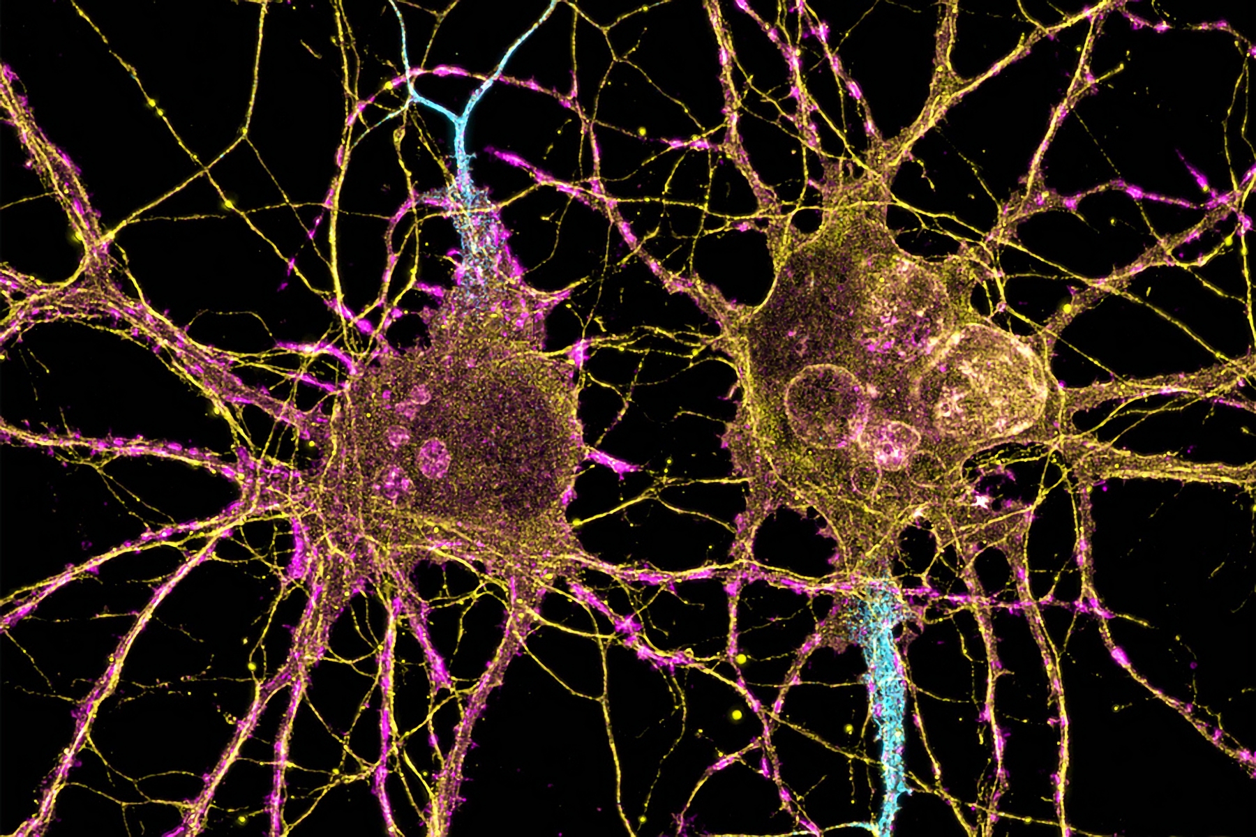
| Neuroscientists at MIT have shown that two distinct cell populations in the striatum are affected differently by Huntington’s disease. Image Credit: Leterrier, NeuroCyto Lab, INP, Marseille, France |
In patients with Huntington’s disease, neurons in a part of the brain called the striatum are among the hardest-hit. Degeneration of these neurons contributes to patients’ loss of motor control, which is one of the major hallmarks of the disease.
Neuroscientists at MIT have now shown that two distinct cell populations in the striatum are affected differently by Huntington’s disease. They believe that neurodegeneration of one of these populations leads to motor impairments, while damage to the other population, located in structures called striosomes, may account for the mood disorders that are often see in the early stages of the disease.
“As many as 10 years ahead of the motor diagnosis, Huntington’s patients can experience mood disorders, and one possibility is that the striosomes might be involved in these,” says Ann Graybiel, an MIT Institute Professor, a member of MIT’s McGovern Institute for Brain Research, and one of the senior authors of the study.
Using single-cell RNA sequencing to analyze the genes expressed in mouse models of Huntington’s disease and postmortem brain samples from Huntington’s patients, the researchers found that cells of the striosomes and another structure, the matrix, begin to lose their distinguishing features as the disease progresses. The researchers hope that their mapping of the striatum and how it is affected by Huntington’s could help lead to new treatments that target specific cells within the brain.
.jpg)


.jpg)

.jpg)


.jpg)


.jpg)