. Scientific Frontline

Tuesday, September 27, 2022

Revealing the Genome of the Common Ancestor of All Mammals

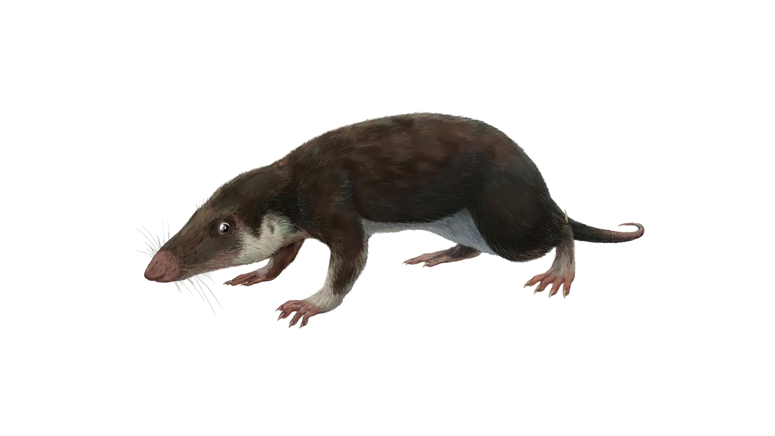
An international team has reconstructed the genome organization of the earliest common ancestor of all mammals. The reconstructed ancestral genome could help in understanding the evolution of mammals and in conservation of modern animals. The earliest mammal ancestor likely looked like this fossil animal, Morganucodon, which lived about 200 million years ago.
Image via Wikipedia by user Funkmonk, Creative Commons Attribution-Share Alike 3.0 Unported license.

Every modern mammal, from a platypus to a blue whale, is descended from a common ancestor that lived about 180 million years ago. We don’t know a great deal about this animal, but the organization of its genome has now been computationally reconstructed by an international team of researchers. The work is published in Proceedings of the National Academy of Sciences.

“Our results have important implications for understanding the evolution of mammals and for conservation efforts,” said Harris Lewin, distinguished professor of evolution and ecology at the University of California, Davis, and senior author on the paper.

The researchers drew on high-quality genome sequences from 32 living species representing 23 of the 26 known orders of mammals. They included humans and chimps, wombats and rabbits, manatees, domestic cattle, rhinos, bats and pangolins. The analysis also included the chicken and Chinese alligator genomes as comparison groups. Some of these genomes are being produced as part of the Earth BioGenome Project and other large-scale biodiversity genome sequencing efforts. Lewin chairs the Working Group for the Earth BioGenome Project.

Novel imaging system could mean near-instant biopsy results

Tissue biopsied with a novel imaging system based on 2-photon fluorescence microscopy (TPFM) is showing promising results. The system, described in the journal JAMA Dermatology, was developed by University of Rochester biomedical engineer Michael Giacomelli.
Photo credit: Giacomelli lab

Medicine has advanced dramatically during the last century. But when it comes to getting biopsy results, very little has changed. Consider, for example, what happens when a patient comes in to have a skin lesion biopsied for nonmelanoma skin cancer.

“The surgeon will take a little piece of the skin out,” says Michael Giacomelli, an assistant professor of biomedical engineering and of optics at the University of Rochester. “Someone in pathology will look at it weeks or even a month later under a microscope. And then, depending on what they find, the patient is notified that everything’s fine, don’t worry about it, or we need you to come back for a second appointment so we can treat you.”

Giacomelli is developing a novel imaging system, contained on a portable cart, to shorten this process to two minutes. This would enable a surgeon to immediately determine whether the lesion is cancerous and, if so, to “treat the patient during the same visit instead of stretching it out over the next month and multiple visits.”

The system—using two-photon fluorescence microscopy (TPFM)—demonstrated remarkable accuracy in a pilot study summarized recently in JAMA Dermatology. When tested on 15 biopsies of known nonmelanoma skin cancer, the technology was able to detect basal cell carcinoma with perfect accuracy (100 percent sensitivity and specificity) and squamous cell carcinoma with high accuracy (89 percent sensitivity and 100 percent specificity).

Seawater could have provided phosphorous required for emerging life

Artist Concept of an Early Earth 
Credit: NASA

The problem of how phosphorus became a universal ingredient for life on Earth may have been solved by researchers from the University of Cambridge and the University of Cape Town, who have recreated primordial seawater containing the element in the lab.

Their results, published in the journal Nature Communications, show that seawater might be the missing source of phosphate, meaning that it could have been available on a large enough scale for life without requiring special environmental conditions.

“This could really change how we think about the environments in which life first originated,” said co-author Professor Nick Tosca from Cambridge's Department of Earth Sciences.

The study, which was led by Matthew Brady, a PhD student from Cambridge's Department of Earth Sciences, shows that early seawater could have held one thousand to ten thousand times more phosphate than previously estimated — as long as the water contained a lot of iron.

Phosphate is an essential ingredient in creating life’s building blocks — forming a key component of DNA and RNA — but it is one of the least abundant elements in the cosmos in relation to its biological importance. When in its mineral form, phosphate is also relatively inaccessible — it can be hard to dissolve in water so that life can use it.

Magnetic Field Helps Thick Battery Electrodes Tackle Electric Vehicle Challenges

Source: University of Texas at Austin
As electric vehicles grow in popularity, the spotlight shines more brightly on some of their remaining major issues. Researchers at The University of Texas at Austin are tackling two of the bigger challenges facing electric vehicles: limited range and slow recharging.

The researchers fabricated a new type of electrode for lithium-ion batteries that could unleash greater power and faster charging. They did this by creating thicker electrodes – the positively and negatively charged parts of the battery that deliver power to a device – using magnets to create a unique alignment that sidesteps common problems associated with sizing up these critical components.

The result is an electrode that could potentially facilitate twice the range on a single charge for an electric vehicle, compared with a battery using an existing commercial electrode.

“Two-dimensional materials are commonly believed as a promising candidate for high-rate energy storage applications because it only needs to be several nanometers thick for rapid charge transport,” said Guihua Yu, a professor in UT Austin’s Walker Department of Mechanical Engineering and Texas Materials Institute. “However, for thick-electrode-design-based next-generation, high-energy batteries, the restacking of nanosheets as building blocks can cause significant bottlenecks in charge transport, leading to difficulty in achieving both high energy and fast charging.”

The key to the discovery, published in the Proceedings of the National Academy of Sciences, uses thin two-dimensional materials as the building blocks of the electrode, stacking them to create thickness and then using a magnetic field to manipulate their orientations. The research team used commercially available magnets during the fabrication process to arrange the two-dimensional materials in a vertical alignment, creating a fast lane for ions to travel through the electrode.

To Stop Viruses, SDSU Researchers are Figuring Out How They're Built

Multiple protein subunits (green, purple and red) of a plant-infecting virus have separate nucleation and growth phases similar to the MS2 bacteria-infecting virus (right).
Source: Protein Data Bank.

An SDSU team, along with Harvard and UCLA collaborators, are researching how distantly related viruses self-organize to improve disease-fighting tactics.

Without a multi-page instruction manual or a commanding Captain America, how do viruses assemble hundreds of individual pieces into elaborate structures capable of spreading disease?

Solving the secret of self-assembly can pave the way for engineering advancements like molecules and robots that put themselves together. It could also contribute to more efficient packaging, automated delivery and targeted design of medicine in the fight against viruses that cause colds, diarrhea, liver cancer and polio.

“If we understand the physical rules of how viruses assemble, then we can try to make them form incorrect structures to hinder their spread,” said Rees Garmann, a chemist at San Diego State University and lead author of a new paper published in the journal PNAS that fills in a piece of the puzzle.

An ocean inside the Earth? Water hundreds of kilometers down

The diamond from Botswana revealed to the scientists that considerable amounts of water are stored in the rock at a depth of more than 600 kilometers.
Photo credit: Tingting Gu, Gemological Institute of America, New York, NY, USA

The transition zone between the Earth's upper and lower mantle contains considerable quantities of water, according to an international study involving the Institute for Geosciences at Goethe University in Frankfurt. The German-Italian-American research team analyzed a rare diamond formed 660 meters below the Earth's surface using techniques including Raman spectroscopy and FTIR spectrometry. The study confirmed something that for a long time was only a theory, namely that ocean water accompanies subducting slabs and thus enters the transition zone. This means that our planet's water cycle includes the Earth's interior. 

Study published in the journal Nature Geoscience.

An ocean inside the Earth? Water hundreds of kilometers down It is located at a depth of 410 to 660 kilometers. The immense pressure of up to 23,000 bar in the TZ causes the olive-green mineral olivine, which constitutes around 70 percent of the Earth's upper mantle and is also called peridot, to alter its crystalline structure. At the upper boundary of the transition zone, at a depth of about 410 kilometers, it is converted into denser wadsleyite; at 520 kilometers it then metamorphoses into even denser ringwoodite.

Saturday, September 24, 2022

Is SARS-CoV-2 hiding in your fat cells?

Left: Catherine Blish, MD, PhD, professor of infectious diseases. Right: Tracey McLaughlin, MD, professor of endocrinology.
Source: Stanford Medicine | Stanford University

A study by Stanford Medicine investigators shows that SARS-CoV-2 can infect human fat tissue. This phenomenon was seen in laboratory experiments conducted on fat tissue excised from patients undergoing bariatric and cardiac surgeries, and later infected in a laboratory dish with SARS-CoV-2. It was further confirmed in autopsy samples from deceased COVID-19 patients.

Obesity is an established, independent risk factor for SARS-CoV-2 infection as well as for the patients’ progression, once infected, to severe disease and death. Reasons offered for this increased vulnerability range from impaired breathing resulting from the pressure of extra weight to altered immune responsiveness in obese people.

But the new study provides a more direct reason: SARS-CoV-2, the virus that causes COVID-19, can directly infect adipose tissue (which most of us refer to as just plain “fat”). That, in turn, cooks up a cycle of viral replication within resident fat cells, or adipocytes, and causes pronounced inflammation in immune cells that hang out in fat tissue. The inflammation even converts uninfected “bystander” cells within the tissue into an inflammatory state.

Discovery of Er Blood Group System


Scientists from the University of Bristol and NHS Blood & Transplant (NHSBT) have discovered a rare new blood group system. The findings, published in Blood, the journal of the American Society of Hematology, also solve a 30-year mystery.

A person’s blood type is determined by the presence or absence of proteins known as blood groups that are present on the surface of red blood cells. Although most people are familiar with the concept of blood groups such as ABO or Rh (the plus or minus), there are many other important blood groups. Where mismatches exist between one person’s blood and that of another, the possibility of alloimmunization (the process by which a person generates an antibody against a blood group antigen that they do not carry) arises. The presence of alloantibodies can have clinical consequences in transfusion or pregnancy by triggering an attack by the immune system

Researchers from Bristol’s School of Biochemistry and NHSBT’s International Blood Group Reference Laboratory (IBGRL) spearheaded an international collaboration which sought to investigate a 30-year mystery surrounding the basis of three known, but genetically uncharacterized, antigens that did not fit into any known blood group system.

Friday, September 23, 2022

A potential new treatment for brain tumors

Featured photo at top of Pankaj Desai, left, and senior graduate research assistant Aniruddha Karve, right, in the lab.
Photo credit: Andrew Higley | University of Cincinnati

A research question posed in Pankaj Desai’s lab has led to a decade of research, a clinical trial and major national funding to further investigate a potential new treatment for the deadliest form of brain tumors.

Desai, PhD, and his team at the University of Cincinnati recently received a $1.19 million grant from the National Institutes of Health/National Institute of Neurological Disorders and Stroke to continue research into the use of a drug called letrozole to treat glioblastomas (GBM).

Research progression

GBMs are aggressive brain tumors that patients often are unaware of until symptoms emerge and the tumor is substantial. Current treatments include immediate surgery to safely remove as much tumor as possible, radiation and chemotherapy, but the tumor often recurs or becomes resistant to treatments. The average patient survives no more than 15 months after diagnosis.

The medication letrozole was approved by the U.S. Food and Drug Administration as a treatment for postmenopausal women with breast cancer in 2001. The drug works by targeting an enzyme called aromatase that is present in breast cancer cells and helps the cancer grow.

DNA nets capture COVID-19 virus in low-cost rapid-testing platform

Tiny nets woven from DNA strands cover the spike proteins of the virus that causes COVID-19 and give off a glowing signal in this artist’s rendering. 
Image courtesy of Xing Wang

Tiny nets woven from DNA strands can ensnare the spike protein of the virus that causes COVID-19, lighting up the virus for a fast-yet-sensitive diagnostic test – and also impeding the virus from infecting cells, opening a new possible route to antiviral treatment, according to a new study.

Researchers at the University of Illinois Urbana-Champaign and collaborators demonstrated the DNA nets’ ability to detect and impede COVID-19 in human cell cultures in a paper published in the Journal of the American Chemical Society.

“This platform combines the sensitivity of clinical PCR tests and the speed and low cost of antigen tests,” said study leader Xing Wang, a professor of bioengineering and of chemistry at Illinois. “We need tests like this for a couple of reasons. One is to prepare for the next pandemic. The other reason is to track ongoing viral epidemics – not only coronaviruses, but also other deadly and economically impactful viruses like HIV or influenza.”

DNA is best known for its genetic properties, but it also can be folded into custom nanoscale structures that can perform functions or specifically bind to other structures much like proteins do. The DNA nets the Illinois group developed were designed to bind to the coronavirus spike protein – the structure that sticks out from the surface of the virus and binds to receptors on human cells to infect them. Once bound, the nets give off a fluorescent signal that can be read by an inexpensive handheld device in about 10 minutes.

Featured Article

Discovery of unexpected collagen structure could ‘reshape biomedical research’

Jeffrey Hartgerink is a professor of chemistry and bioengineering at Rice. Photo Credit: Courtesy of Jeffrey Hartgerink / Rice University Co...

Top Viewed Articles