. Scientific Frontline

Monday, January 3, 2022

3D semiconductor particles offer 2D properties

When it comes to creating next-generation electronics, two-dimensional semiconductors have a big edge. They’re faster, more powerful and more efficient. They’re also incredibly difficult to fabricate.

Three-dimensional semiconductor particles have an edge, too – many of them – given their geometrically varied surfaces. Cornell researchers have discovered that the junctures at these facet edges have 2D properties, which can be leveraged for photoelectrochemical processes – in which light is used to drive chemical reactions – that can boost solar energy conversion technologies.

This research, led by Peng Chen, the Peter J.W. Debye Professor of Chemistry in the College of Arts and Sciences, could also benefit renewable energy technologies that reduce carbon dioxide, convert nitrogen into ammonia, and produce hydrogen peroxide.

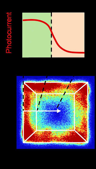
A high-resolution map of a photocatalyst particle shows the transition zones of reactivity and the corresponding spatial variation of photoelectrochemical performance across the inter-facet edge.

The group’s paper, “Inter-Facet Junction Effects on Particulate Photoelectrodes,” published in Nature Materials. The paper’s lead author is postdoctoral researcher Xianwen Mao.

For their study, the researchers focused on the semiconductor bismuth vanadate, particles of which can absorb light and then use that energy to oxidize water molecules – a clean way of generating hydrogen as well as oxygen.

The semiconductor particles themselves are anisotropically-shaped; that is, they have 3D surfaces, full of facets angled toward each other and meeting at edges on the particle surface. However, not all facets are equal. They can have different structures that, in turn, result in different energy levels and electronic properties.

T cells fit to tackle Omicron, suggests

A transmission electron micrograph of negative-stained SARS-CoV-2 Omicron variant virions,
false-colored.
Image: Dr Jason A. Roberts, Head of Electron Microscopy and Structural Virology at The Royal Melbourne Hospital's Victorian Infectious Diseases Reference Laboratory, Doherty Institute

Research from the University of Melbourne and Hong Kong University of Science and Technology (HKUST) has revealed T cells, one of the body’s key defenses against COVID-19, are expected to be effective in mounting an immune response against Omicron despite its significantly higher mutations compared to previous variants of concern.

T cells, generated both by vaccinations and COVID-19 infections, have been shown to be critical in limiting progression to severe disease by eliminating virus-infected cells and helping with other immune system functions.

Preliminary studies have reported that Omicron (fast becoming the most dominant circulating strain globally) can escape antibodies produced by vaccination or natural COVID-19 infection, raising concerns about the increased possibility of reinfection and breakthrough cases.

Published in Viruses, the team analyzed over 1,500 fragments of SARS-CoV-2’s viral proteins, called epitopes, that have been found to be recognized by T cells in recovered COVID-19 patients or after vaccination. The team’s findings suggest Omicron is unlikely to be able to evade T cells, adding to a growing body of evidence from research groups around the world who are also investigating T cell responses to COVID-19.

Secondary structures in DNA are associated with cancer

A new cancer study reports that DNA manifested as knot-like folds and third rungs between DNA’s two strands may drive cancer development and an important regulatory enzyme could be associated with the formation of these unusual structures.

Scientists from Northwestern Medicine and the La Jolla Institute for Immunology (LJI) have discovered that the loss of TET enzymes — a family of enzymes crucial for removing DNA methylation marks — is associated with B-cell lymphoma. Reduced activity of TET enzymes is common in many different cancers. Understanding the mechanisms behind cancer development upon loss of TET function may open the door for new drug treatment strategies to target multiple cancers.

The research was recently published in the journal Nature Immunology.

Previous research demonstrated specific mutations in cancer cells can result in loss of TET function in patients with blood cancers and solid cancers, causing delays in cell communication. Past studies have also identified genomic instabilities such as double-stranded breaks in the DNA code in cancer cells.

Before now, the two dangerous cell features had not been linked.

Strange, unusual structures appear in the DNA

Vipul Shukla, an assistant professor of cell and developmental biology at Northwestern University Feinberg School of Medicine, along with Anjana Rao, a professor in LJI Center for Cancer Immunotherapy, and Daniela Samaniego-Castruita, a University of California San Diego graduate student, hoped to explore one potential way TET deficiency and genomic instability may be connected.

Thursday, December 30, 2021

Elusive atmospheric molecule produced in a lab for the 1st time

Methanediol molecule
Credit: University of Hawaiʻi

The previously elusive methanediol molecule of importance to the organic, atmospheric science and astrochemistry communities has been synthetically produced for the first time by University of Hawaiʻi at Mānoa researchers. Their discovery and methods were published in Proceedings of the National Academy of Sciences on December 30.

Methanediol is also known as formaldehyde monohydrate or methylene glycol. With the chemical formula CH2(OH)2, it is the simplest geminal diol, a molecule which carries two hydroxyl groups (OH) at a single carbon atom. These organic molecules are suggested as key intermediates in the formation of aerosols and reactions in the ozone layer of the atmosphere.

The research team—consisting of Department of Chemistry Professor Ralf Kaiser, postdoctoral researchers Cheng Zhu, N. Fabian Kleimeier and Santosh Singh, and W.M. Keck Laboratory in Astrochemistry Assistant Director Andrew Turner—prepared methanediol via energetic processing of extremely low temperature ices and observed the molecule through a high-tech mass spectrometry tool exploiting tunable vacuum photoionization (the process in which an ion is formed from the interaction of a photon with an atom or molecule) in the W.M. Keck Laboratory in Astrochemistry. Electronic structure calculations by University of Mississippi Associate Professor Ryan Fortenberry confirmed the gas phase stability of this molecule and demonstrated a pathway via reaction of electronically excited oxygen atoms with methanol.

Leveraging Space to Advance Stem Cell Science and Medicine

Arun Sharma, PhD, leads a new research laboratory in the Cedars-Sinai Board of Governors Regenerative Medicine Institute, Smidt Heart Institute and Department of Biomedical Sciences.
Photo by Cedars-Sinai.

The secret to producing large batches of stem cells more efficiently may lie in the near-zero gravity conditions of space. Scientists at Cedars-Sinai have found that microgravity has the potential to contribute to life-saving advances on Earth by facilitating the rapid mass production of stem cells.

A new paper, led by Cedars Sinai and published in the peer-reviewed journal Stem Cell Reports, highlights key opportunities discussed during the 2020 Biomanufacturing in Space Symposium to expand the manufacture of stem cells in space.

Biomanufacturing—a type of stem cell production that uses biological materials such as microbes to produce substances and biomaterials suitable for use in preclinical, clinical, and therapeutic applications—can be more productive in microgravity conditions.

“We are finding that spaceflight and microgravity is a desirable place for biomanufacturing because it confers a number of very special properties to biological tissues and biological processes that can help mass produce cells or other products in a way that you wouldn’t be able to do on Earth,” said stem cell biologist Arun Sharma, PhD, research scientist and head of a new research laboratory in the Cedars-Sinai Board of Governors Regenerative Medicine Institute, Smidt Heart Institute and Department of Biomedical Sciences.

High-resolution lab experiments show how cells ‘eat’

Comert Kural
A new study shows how cell membranes curve to create the “mouths” that allow the cells to consume things that surround them.

“Just like our eating habits basically shape anything in our body, the way cells ‘eat’ matters for the health of the cells,” said Comert Kural, associate professor of physics at The Ohio State University and lead author of the study. “And scientists did not, until now, understand the mechanics of how that happened.”

The study, published recently in the journal Developmental Cell, found that the intercellular machinery of a cell assembles into a highly curved basket-like structure that eventually grows into a closed cage. Scientists had previously believed that structure began as a flat lattice.

Membrane curvature is important, Kural said: It controls the formation of the pockets that carry substances into and out of a cell.

The pockets capture substances around the cell, forming around the extracellular substances, before turning into vesicles – small sacs one-one millionth the size of a red blood cell. Vesicles carry important things for a cell’s health – proteins, for example – into the cell. But they can also be hijacked by pathogens that can infect cells.

But the question of how those pockets formed from membranes that were previously believed to be flat had stymied researchers for nearly 40 years.

“It was a controversy in cellular studies,” Kural said. “And we were able to use super-resolution fluorescence imaging to actually watch these pockets form within live cells, and so we could answer that question of how they are created.

Wednesday, December 29, 2021

Smart sutures to monitor deep surgical wounds

Surgical sutures with an attached electronic module for wireless and battery-free monitoring of deep surgical sites.
Credit: National University of Singapore

Monitoring surgical wounds after an operation is an important step to prevent infection, wound separation and other complications.

However, when the surgical site is deep in the body, monitoring is normally limited to clinical observations or costly radiological investigations that often fail to detect complications before they become life-threatening.

Hard bioelectronic sensors can be implanted in the body for continuous monitoring, but may not integrate well with sensitive wound tissue.

To detect wound complications as soon as they happen, a team of researchers led by Assistant Professor John Ho from the NUS Electrical and Computer Engineering as well as the NUS Institute for Health Innovation & Technology has invented a smart suture that is battery-free and can wirelessly sense and transmit information from deep surgical sites.

These smart sutures incorporate a small electronic sensor that can monitor wound integrity, gastric leakage and tissue micromotions, while providing healing outcomes which are equivalent to medical-grade sutures.

Robots collect underwater litter

The robot of the SeaClear Project is able to detect and collect underwater litter.
Image: The SeaClear Project
Removing litter from oceans and seas is a costly and time-consuming process. As part of a European cooperative project, a team at the Technical University of Munich (TUM) is developing a robotic system that uses machine learning methods to locate and collect waste under water.

Our seas and oceans currently contain somewhere between 26 and 66 million tons of plastic waste, most of which is lying on the seafloor. This represents an enormous threat to marine plants and animals and to the ecological balance of the seas.

But removing waste from the waters is a complex and expensive process. It is often dangerous, too, because the work is generally done by scuba divers. The cleanup operations are also usually limited to the water surface. In the SeaClear Project, a team at TUM is working with eight European partner institutions to develop a robotic system capable of collecting underwater litter.

Tuesday, December 28, 2021

Optimization of mRNA containing nanoparticles

Dr. Aurel Radulescu at the KWS-2 instrument of the Juelich Center for Neutron Science (JCNS) in the research neutron source Heinz Maier-Leibnitz (FRM II) of the Technical University of Munich
Image: Bernhard Ludewig / TUM / FRM II

The research neutron source Hein Maier-Leibnitz (FRM II) at the Technical University of Munich (TUM) is playing an important role in the investigation of mRNA nanoparticles similar to the ones used in the Covid-19 vaccines from vendors BioNTech and Pfizer. Researchers at the Heinz Maier-Leibnitz Zentrum (MLZ) used the high neutron flux available in Garching to characterize various formulations for the mRNA vaccine and thus to lay the groundwork for improving the vaccine's efficacy.

The idea of using messenger RNA (mRNA) as an active ingredient is a brilliant one: The molecule contains the specific blueprint for proteins which are then synthesize by the cell. This makes it generally possible to provide a very wide spectrum of different therapeutically effective proteins.

In the case of the Covid-19 vaccine, these are the proteins of the characteristic spikes on the surface of the Corona virus which are used for vaccination. The proteins are presented on the surface of immune cells; then the human immune system triggers defenses against these foreign proteins and thus against the Corona virus. The mRNA itself is completely broken down after only a few hours, a fact which is advantageous to the safety of these vaccines.

The mRNA has to be packaged appropriately in order to keep it from being broken down on the way to the cell by the ubiquitous enzymes of the human body. This is done using nanoparticles which can consist of a mixture of lipids or polymers.

Engineers bring a soft touch to commercial robotics

Credit: National University of Singapore
Inspired by the natural dexterity of the human hand, a team of engineers from the National University of Singapore (NUS) has created a reconfigurable hybrid robotics system that is able to grip a variety of objects: from the small, soft and delicate to the large, heavy and bulky. This technology is expected to impact a range of industries, involving food assembly, vertical farming and fast-moving consumer goods packaging, which will progressively automate more of their operations in the coming years.

Led by Associate Professor Raye Yeow from the NUS Department of Biomedical Engineering and the NUS Advanced Robotics Centre, the hybrid robotic grippers use soft, flexible 3D-printed fingers with a reconfigurable gripper base. The robotic innovation is now in the process of being brought to commercial partners under the team’s start-up RoPlus (RO+), comprising NUS researchers Low Jin Huat, Khin Phone May, Chen Chao-Yu and undergraduate student Han Qian Qian.

“An object’s shape, texture, weight and size affect how we choose to grip them. This is one of the main reasons why many industries still heavily rely on human labour to package and handle delicate items,” Assoc Prof Yeow said. “Our hybrid robotic gripper technology revolutionizes traditional pick-and-place tasks by offering advanced capabilities that allow robots to safely interact with delicate items of various shapes, sizes and stiffness, just like the human hand.”

Monday, December 27, 2021

Window to access legal abortion may close before many women know they are pregnant

Credit: Heidi-Ann Fourkiller / SFLORG
More than one in five women experience irregular menstrual cycles that could keep them from learning they are pregnant until it may be too late to access an abortion under some state laws in effect or under consideration, a new study shows.

Researchers from the University of Wisconsin–Madison and National Institutes of Health analyzed anonymized data on 1.6 million menstrual cycles provided by more than 267,000 adults to a cycle-tracking app. According to results published today in the journal Proceedings of the National Academy of Sciences, 22 percent of the people in the study had irregular menstrual cycles that differed in length from one cycle to the next by seven or more days.

“For almost everyone, the first symptom of a pregnancy is a missed period,” says UW–Madison sociology Professor Jenna Nobles, coauthor of the study. “But many people — a large share of the population — have long or highly irregular cycles and could not reasonably learn about their pregnancy in time to seek a legal abortion under laws that set limits at detectable fetal cardiac activity or six weeks.”

Irregular periods are more likely among those with some relatively common medical conditions like Type 2 diabetes, polycystic ovary syndrome and thyroid and other hormone disorders. Hispanic women had greater risk than non-Hispanic white women of experiencing irregular cycles. The age group most like to have cycles of irregular length is 18- to 24-year-olds — also the ages with the highest abortion rates in the United States.

‘Battle of the sexes’ begins in womb as father and mother’s genes tussle over nutrition

Section of mouse fetus and placenta 
Credit: Ionel Sandovici
As the fetus grows, it needs to communicate its increasing needs for food to the mother. It receives its nourishment via blood vessels in the placenta, a specialized organ that contains cells from both baby and mother.

Between 10% and 15% of babies grow poorly in the womb, often showing reduced growth of blood vessels in the placenta. In humans, these blood vessels expand dramatically between mid and late gestation, reaching a total length of approximately 320 kilometers at term.

In a study published today in Developmental Cell, a team led by scientists at the University of Cambridge used genetically engineered mice to show how the fetus produces a signal to encourage growth of blood vessels within the placenta. This signal also causes modifications to other cells of the placenta to allow for more nutrients from the mother to go through to the fetus.

Dr Ionel Sandovici, the paper’s first author, said: “As it grows in the womb, the fetus needs food from its mum, and healthy blood vessels in the placenta are essential to help it get the correct amount of nutrients it needs.

“We’ve identified one way that the fetus uses to communicate with the placenta to prompt the correct expansion of these blood vessels. When this communication breaks down, the blood vessels don’t develop properly and the baby will struggle to get all the food it needs.”

Scientists build new atlas of ocean’s oxygen-starved waters

Oxygen deficient zone intensity across the eastern Pacific Ocean, where copper colors represent the locations of consistently lowest oxygen concentrations and deep teal indicates regions without sufficiently low dissolved oxygen.
Credit: Jarek Kwiecinski and Andrew Babbin

Life is teeming nearly everywhere in the oceans, except in certain pockets where oxygen naturally plummets and waters become unlivable for most aerobic organisms. These desolate pools are “oxygen-deficient zones,” or ODZs. And though they make up less than 1 percent of the ocean’s total volume, they are a significant source of nitrous oxide, a potent greenhouse gas. Their boundaries can also limit the extent of fisheries and marine ecosystems.

Now MIT scientists have generated the most detailed, three-dimensional “atlas” of the largest ODZs in the world. The new atlas provides high-resolution maps of the two major, oxygen-starved bodies of water in the tropical Pacific. These maps reveal the volume, extent, and varying depths of each ODZ, along with fine-scale features, such as ribbons of oxygenated water that intrude into otherwise depleted zones.

The team used a new method to process over 40 years’ worth of ocean data, comprising nearly 15 million measurements taken by many research cruises and autonomous robots deployed across the tropical Pacific. The researchers compiled then analyzed this vast and fine-grained data to generate maps of oxygen-deficient zones at various depths, similar to the many slices of a three-dimensional scan.

Sunday, December 26, 2021

Helium bath splash

Ions packed in a helium nanodroplet remain protected on impact.
Credit: University Innsbruck

While working with helium nanodroplets, scientists at the Department of Ion Physics and Applied Physics led by Fabio Zappa and Paul Scheier have come across a surprising phenomenon: When the ultracold droplets hit a hard surface, they behave like drops of water. Ions with which they were previously doped thus remain protected on impact and are not neutralized.

At the Department of Ion Physics and Applied Physics, Paul Scheier's research group has been using helium nanodroplets to study ions with methods of mass spectrometry for around 15 years. Using a supersonic nozzle, tiny, superfluid helium nanodroplets can be produced with temperatures of less than one degree Kelvin. They can very effectively be doped with atoms and molecules. In the case of ionized droplets, the particles of interest are attached to the charges, which are then measured in the mass spectrometer. During their experiments, the scientists have now stumbled upon an interesting phenomenon that has fundamentally changed their work. "For us, this was a gamechanger," says Fabio Zappa from the nano-bio-physics team. "Everything at our lab is now done with this newly discovered method." The researchers have now published the results of their studies in Physical Review Letters.

Saturday, December 25, 2021

Schizophrenia and bipolar disorder found in recently evolved region of the ‘dark genome’

They say these new proteins can be used as biological indicators to distinguish between the two conditions, and to identify patients more prone to psychosis or suicide.

Schizophrenia and bipolar disorder are debilitating mental disorders that are hard to diagnose and treat. Despite being amongst the most heritable mental health disorders, very few clues to their cause have been found in the sections of our DNA known as genes.

The scientists think that hotspots in the ‘dark genome’ associated with the disorders may have evolved because they have beneficial functions in human development, but their disruption by environmental factors leads to susceptibility to, or development of, schizophrenia or bipolar disorder.

The results are published in the journal Molecular Psychiatry.

“By scanning through the entire genome we’ve found regions, not classed as genes in the traditional sense, which create proteins that appear to be associated with schizophrenia and bipolar disorder,” said Dr Sudhakaran Prabakaran, who was based in the University of Cambridge’s Department of Genetics when he conducted the research, and is senior author of the report.

He added: “This opens up huge potential for new druggable targets. It’s really exciting because nobody has ever looked beyond the genes for clues to understanding and treating these conditions before.”

The researchers think that these genomic components of schizophrenia and bipolar disorder are specific to humans - the newly discovered regions are not found in the genomes of other vertebrates. It is likely that the regions evolved quickly in humans as our cognitive abilities developed, but they are easily disrupted - resulting in the two conditions.

Featured Article

What Is: Psychopathy | Part three of the "Dark Tetrad"

Scientific Frontline: Extended "At a Glance" Summary : Psychopathy The Core Concept : Psychopathy is a profound personality disord...

Top Viewed Articles