. Scientific Frontline

Friday, September 30, 2022

Chipping away at the many unknowns of obscure animal viruses

Patas monkeys are among the wild African monkeys believed to be natural reservoirs for the simian hemorrhagic fever virus. 
Photo Credit: Andrew S

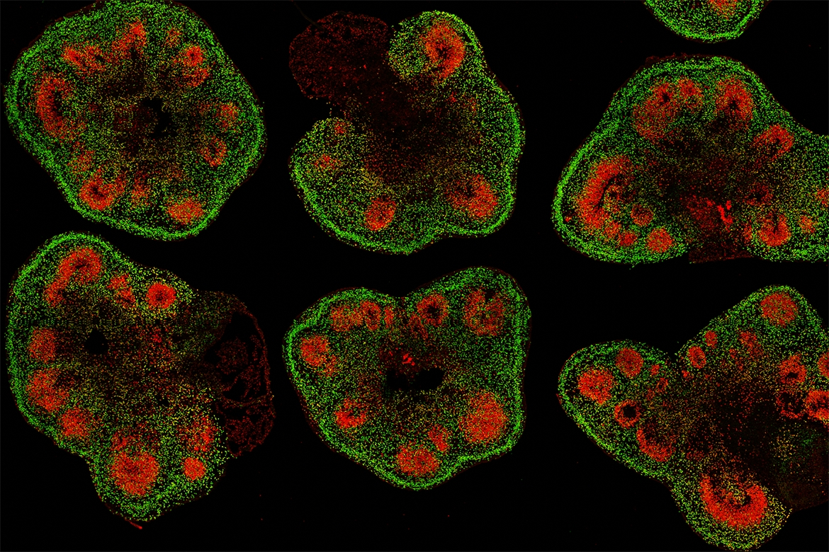
Researchers have identified enough biological details about a virus endemic in African primates to suggest that this virus, which causes a hemorrhagic fever disease in monkeys, has decent potential to spill over to humans.

The findings suggest a surveillance program is warranted for citizens in Africa who may be at risk for exposure to the virus. But the study teaches a much larger lesson as well, researchers say: It’s never too early to start preparing for the next animal virus to come along and unexpectedly cause disease in people.

“There are a lot of unknown animal viruses out there that may pose risk to humans,” said Cody Warren, first author of the study and assistant professor of veterinary biosciences at The Ohio State University.

“We need to be prospectively looking at animal viruses that have been ignored to see if they have the capacity to replicate in human cells. If they do, will we continue to ignore them? I don’t think we should,” he said.

Warren completed this work at the University of Colorado Boulder as a postdoctoral researcher in the lab of senior author Sara Sawyer, professor of molecular, cellular & developmental biology.

Research finds link between poor health and low breast milk production

Photo Credit: seeseehundhund

Research from the University of Cincinnati shows that poor metabolic health parameters are linked to low breast milk production. The study was published in the journal Breastfeeding Medicine.

“We wanted to see if we could understand what stands out as different in these moms. So, we conducted a case control study to see why, despite their best efforts at doing everything right with breastfeeding, they were not making enough milk,” says Laurie Nommsen-Rivers, PhD, associate professor of nutrition, and the Ruth Rosevear Endowed Chair of Maternal and Child Nutrition in the UC College of Allied Health Sciences. “The prevailing dogma is if you try hard enough at breastfeeding, your body will be able to do this.”

Nommsen-Rivers and her team analyzed data from a randomized controlled trial from February 2015 to June 2016 involving women screened for a low-milk supply. Mothers who were aged 20 years or older and one to eight weeks postpartum with a healthy infant born at 37 weeks of gestation or later were included. Participants completed at-home infant test-weighing to measure milk output.

New Superconducting Qubit Testbed Benefits Quantum Information Science Development

A superconducting qubit sits in a dilution refrigerator in a Pacific Northwest National Laboratory (PNNL) physics lab. This experimental device is the first step in establishing a qubit testbed at PNNL.
  Photo Credit: Andrea Starr | Pacific Northwest National Laboratory

If you’ve ever tried to carry on a conversation in a noisy room, you’ll be able to relate to the scientists and engineers trying to “hear” the signals from experimental quantum computing devices called qubits. These basic units of quantum computers are early in their development and remain temperamental, subject to all manner of interference. Stray “noise” can masquerade as a functioning qubit or even render it inoperable.

That’s why physicist Christian Boutan and his Pacific Northwest National Laboratory (PNNL) colleagues were in celebration mode recently as they showed off PNNL’s first functional superconducting qubit. It’s not much to look at. Its case—the size of a pack of chewing gum--is connected to wires that transmit signals to a nearby panel of custom radiofrequency receivers. But most important, it’s nestled within a shiny gold cocoon called a dilution refrigerator and shielded from stray electrical signals. When the refrigerator is running, it is among the coldest places on Earth, so very close to absolute zero, less than 6 millikelvin (about −460 degrees F).

Stone spheres could be from Ancient Greek board game

Groups of spheres from Akrotiri
Photo Credit: Konstantinos Trimmis

Scientific Frontline: "At a Glance" Summary

  • Main Discovery: Archaeologists from the University of Bristol have determined that mysterious stone spheres found in ancient Aegean settlements, specifically Akrotiri, are likely playing pieces from one of the earliest known board games.
  • Methodology: The research team analyzed 700 stone spheres dating between 4,500 and 3,600 years old, examining specific features such as size, material, and color to identify usage patterns.
  • Key Statistic: The analysis revealed that the stones consistently fit into two distinct size clusters (large and small), contradicting the multiple groupings that would be expected if they were used for a counting or record-keeping system.
  • Context: This hypothesis aligns the artifacts with similar ancient board games from the Levant and Egypt, such as Mehen and Senet, replacing previous theories that the stones were used for slings or tossing.
  • Significance: The specific deposition of the spheres in stone cavities (kernos) indicates they were objects of social importance, providing new evidence of social interaction and leisure in the Bronze Age Aegean.
  • Future Application: Researchers plan to utilize artificial intelligence techniques to determine the actual gameplay mechanics and will apply clustering analysis to the associated stone slabs to verify the link.

Container for Hazardous Radioactive Waste Storage Model Created

According to Oleg Tashlykov, the container protects from radiation in all directions.
Photo Credit: Anastasia Farafontova

Ural Federal University scientists designed a container to store solidified liquid radioactive waste containing "long-lived" cesium-137 and cobalt-60, the most potentially dangerous of all radioactive waste. Due to their innovative design and filling, the simulated containers are capable of reducing radiation from radioactive waste to safe levels. One such container could replace five or six of the standard type. An article about the scientific work was published in the journal Progress in Nuclear Energy.

The modeled container consists of three main layers: a stainless steel inner capsule, halloysite clay filler, and an outer cementation concrete layer. The stainless steel capsule holds more than 450,000 cm3 of radioactive waste. Radionuclides are concentrated in a special sorbent, which is used in ion-selective purification and is placed inside the capsule. Stainless steel was chosen because, unlike carbon steel, it is more resistant to corrosion and does not require shielding.

"As a rule, such containers consist of two layers: outer cementation concrete and an inner metallic hosting capsule with a radioactive sorbent (or a sorbent in a cement matrix is placed inside the container). The main disadvantage of such a container arrangement is that their shielding, i.e. protective, capacity is limited. We suggest a three-layer container - with an additional layer between the inner metal capsule and the outer shell. The material that fills this space must be inexpensive and still effectively reduce the gamma radiation emitted by the radioisotopes inside the radioactive waste container. In this case, we investigated the protective properties of the intermediate layer consisting of halloysite - a fine-dispersed nanoscale white clay with a chemical composition rich in aluminum and silicon," says Oleg Tashlykov, Associate Professor at the Department of Nuclear Power Plants and Renewable Energy Sources at UrFU, Head of Research and one of the authors of the article.

Dual-targeting CAR NK cells can prevent cell dysfunction and tumor escape

 Katy Rezvani, M.D., Ph.D
Credit: The University of Texas MD Anderson Cancer Center.

Researchers at The University of Texas MD Anderson Cancer Center have developed a new approach to engineering natural killer (NK) cells with a second chimeric antigen receptor (CAR) to act as a logic gate, requiring two signals to eliminate a target cell. In preclinical studies, these next-generation CAR NK cells improved tumor specificity and enhanced anti-tumor activity by overcoming a process that contributes to NK cell dysfunction and tumor relapse.

This study, published in Nature Medicine, demonstrated that a normal physiological process called trogocytosis contributes to tumor escape and poor responses after CAR NK cell therapy by causing tumor antigen loss, NK cell exhaustion and fratricide — the killing of sibling CAR NK cells.

“We identified a novel mechanism of relapse following CAR NK cell therapy, and we also have developed a strategy to mitigate this process,” said corresponding author Katy Rezvani, M.D., Ph.D., professor of Stem Cell Transplantation & Cellular Therapy. “We engineered CAR NK cells with dual-targeting CARs that are able to ignore tumor antigens on the surface of their sibling NK cells acquired as a result of trogocytosis and selectively eliminate tumor cells.”

Rezvani and Ye Li, M.D., a graduate student in the Rezvani Lab, led the study.

During trogocytosis, surface proteins from a target cell are transferred to the surface of an immune cell, such as an NK cell or T cell, in order to regulate their activity. Using preclinical models, Li and colleagues showed that CAR activation promotes trogocytosis, resulting in the transfer and expression of tumor antigens on CAR NK cells.

Traumatic brain injury ‘remains a major global health problem’ say experts

Photo Credit: Ian Valerio

The report – the 2022 Lancet Neurology Commission – has been produced by world-leading experts, including co-lead author Professor David Menon from the Division of Anesthesia at the University of Cambridge.

 "Over the last decade, large international collaborations have provided important information to improve understanding and care of TBI. However, significant problems remain, especially in low- and middle-income countries"
David Menon

The Commission documents traumatic brain injury (TBI) as a global public health problem, which afflicts 55 million people worldwide, costs over US$400 billion per year, and is a leading cause of injury-related death and disability.

TBI is not only an acute condition but also a chronic disease with long-term consequences, including an increased risk of late-onset neurodegeneration, such as Parkinson’s disease and dementia. Road traffic incidents and falls are the main causes, but while in low- and middle-income countries, road traffic accidents account for almost three times the number of TBIs as falls, in high-income countries falls cause twice the number of TBIs compared to road traffic accidents. These data have clear consequences for prevention.

Over 90% of TBIs are categorized as ‘mild’, but over half of such patients do not fully recover by six months after injury. Improving outcome in these patients would be a huge public health benefit. A multidimensional approach to outcome assessment is advocated, including a focus on mental health and post-traumatic stress disorder. Outcome after TBI is poorer in females compared with males, but reasons for this are not clear.

Molecular chaperones caught in flagrante

For an adequate immune response, it is essential that T lymphocytes recognize infected or degenerated cells. They do so by means of antigenic peptides, which these cells present with the help of specialized surface molecules (MHC I molecules). Using X-ray structure analysis, a research team from Frankfurt has now been able to show how the MHC I molecules are loaded with peptides and how suitable peptides are selected for this purpose.

As task forces of the adaptive immune system, T lymphocytes are responsible for attacking and killing infected or cancerous cells. Such cells, like almost all cells in the human body, present on their surface fragments of all the proteins they produce inside. If these include peptides that a T lymphocyte recognizes as foreign, the lymphocyte is activated and kills the cell in question. It is therefore important for a robust T-cell response that suitable protein fragments are presented to the T lymphocyte. The research team led by Simon Trowitzsch and Robert Tampé from the Institute of Biochemistry at Goethe University Frankfurt has now shed light on how the cell selects these protein fragments or peptides.

Peptide presentation takes place on so-called major histocompatibility complex class I molecules (MHC I). MHC I molecules are a group of very diverse surface proteins that can bind myriads of different peptides. They are anchored in the cell membrane and form a peptide-binding pocket with their outward-facing part. Like all surface proteins, MHC I molecules take the so-called secretory pathway: they are synthesized into the cell's cavity system (endoplasmic reticulum (ER) and Golgi apparatus) and folded there. Small vesicles then bud off from the cavity system, migrate to the cell membrane and fuse with it.

Study reveals how COVID-19 damages the heart

Image Credit: Sanjay k j

University of Queensland researchers have discovered how COVID-19 damages the heart, opening the door to future treatments.

This initial study – featuring a small cohort – found COVID-19 damaged the DNA in cardiac tissue, which wasn’t detected in influenza samples.

UQ Diamantina Institute researcher Dr Arutha Kulasinghe said the team found while COVID-19 and influenza are both severe respiratory viruses, they appeared to affect cardiac tissue very differently.

“In comparison to the 2009 flu pandemic, COVID has led to more severe and long-term cardiovascular disease but what was causing that at a molecular level wasn’t known,” Dr Kulasinghe said.

“During our study, we couldn’t detect viral particles in the cardiac tissues of COVID-19 patients, but what we found was tissue changes associated with DNA damage and repair.

“DNA damage and repair mechanisms foster genomic instability and are related to chronic diseases such as diabetes, cancer, atherosclerosis and neurodegenerative disorders, so understanding why this is happening in COVID-19 patients is important.”

Paleontologists Found Mammoth Baby, Ancient Bear Teeth, and Lair of Cave Hyenas

Scientists will open a new expedition season in spring.
Photo credit: TASS-Ural Press Center / Vladislav Burnashev

Paleontologists of Ural Federal University and the Institute of Plant and Animal Ecology of Ural Branch of Russian Academy of Sciences during summer expeditions found a large number of ancient bones, teeth, as well as wool and skin of a mammoth baby. The study of remains will allow us to recreate the specifics of the flora and fauna of ancient times in detail and to understand the specifics of animal nutrition. Scientists told about the results of summer expeditions at a press conference in TASS.

On the Gyda Peninsula (Far North), paleontologists found the well-preserved remains of a mammoth baby. The uniqueness of the discovery is its age - it is a six-year-old mammoth baby. If previously only single bones were found, now the researchers have found material that will help study mammoth babies, said Pavel Kosintsev, a leading expert of the Laboratory of Natural Science Methods in Humanities of UrFU, a Senior Researcher of the Institute of Plant and Animal Ecology of the Ural Branch of the Russian Academy of Sciences.

Thursday, September 29, 2022

Ancient 'Shark' from China Is Humans' Oldest Jawed Ancestor

Life reconstruction of Fanjingshania renovata.
Image Credit: ZHANG Heming)

Paleontologists discover a 439-million-year-old 'shark' that forces us to rethink the timeline of vertebrate evolution

Living sharks are often portrayed as the apex predators of the marine realm. Paleontologists have been able to identify fossils of their extinct ancestors that date back hundreds of millions of years to a time known as the Palaeozoic period. These early "sharks," known as acanthodians, bristled with spines. In contrast to modern sharks, they developed bony "armor" around their paired fins.

A recent discovery of a new species of acanthodian from China surprised scientists with its antiquity. The find predates by about 15 million years the earliest acanthodian body fossils and is the oldest undisputed jawed fish.

These findings were published in Nature.

Reconstructed from thousands of tiny skeletal fragments, Fanjingshania, named after the famous UNESCO World Heritage Site Fanjingshan, is a bizarre fish with an external bony "armor" and multiple pairs of fin spines that set it apart from living jawed fish, cartilaginous sharks and rays, and bony ray- and lobe-finned fish.

Making lab-grown brain organoids ‘brainier

 Slices of mini–brain organoids with neural stem cells (red) and cortical neurons (green).
Credit: Hajime Ozaki, Watanabe lab/UCI

By using stem cells to grow miniature brain-like organs in the lab, scientists have opened a new avenue for studies of neurological development, disease and therapies that can’t be conducted in living people. But not all mini–brain organoids are created equal and getting them to precisely mimic the human brain tissues they’re modeling has been a persistent challenge.

“Right now, it’s like the Wild West because there is no standard method for generating mini–brain organoids,” said Bennett Novitch, a member of the Eli and Edythe Broad Center of Regenerative Medicine and Stem Cell Research at UCLA and the senior author of a new paper on the topic. “Every neuroscientist wants to make a brain organoid model of their favorite disease, and yet everyone’s organoids do not always look alike.”

In fact, because there is no common protocol for their production and a lack of quality-control guidelines, organoids can vary from lab to lab — and even from batch to batch — which means that a finding made in one organoid may not hold true in another.

“If my lab and another lab down the hall were to conduct drug screens using mini–brain organoid models of the same disorder, we could still get different results,” said Momoko Watanabe, the new paper’s first author and an assistant professor of anatomy and neurobiology at UC Irvine. “We won’t know whose findings are correct because the differences we’re seeing could be reflections of how our models differ rather than reflections of the disease.”

Wildfire smoke exposure hurts learning outcomes

Exposure to fine particle pollution from wildfire smoke during the school day affects average test scores. In this map of the predicted effect on average test scores by district in a relatively high-smoke year, 2016, darker shades indicate a stronger impact.
Image credit: Wen et al. 2022, Nature Sustainability

Pollution from wildfires is linked to lower test scores and possibly lower future earnings for kids growing up with more smoke days at school, a new study finds. Impacts of smoke exposure on earnings are disproportionately borne by economically disadvantaged communities of color.

When wildfire smoke pollutes the air in schoolyards and classrooms, as it does with increasing frequency and severity across the country, it hurts not only children’s health but also their ability to learn and possibly their future earning power, according to new research from Stanford University.

The new analysis, published Sept. 29 in Nature Sustainability, draws on eight years of standardized test scores from nearly 11,700 public school districts across six grades, as well as estimates of daily smoke exposure derived from satellite measurements.

The researchers found test scores in English language arts and math dropped significantly during school years even at low levels of smoke exposure, and that test-score impacts grew as students’ smoke exposure worsened.

The impact on test scores nearly doubled when students were exposed to heavy smoke during the school day compared to the weekend. Underscoring previous studies suggesting that air pollution impacts are particularly harmful for younger students, the study also revealed greater impacts for third to fifth graders compared to sixth to eighth graders.

WVU engineers bring new life to electronics recycling, address supply chain shortfalls affecting national defense

Edward Sabolsky, WVU Benjamin M. Statler College of Engineering and Mineral Resources professor uses ceramic bricks to conduct research at his lab. The U.S. Department of Defense has tasked Sabolsky and Terence Musho with developing a new process for recycling electronic waste in order to extract raw materials that are used to build technology critical to U.S. national defense, such as semiconductors. Photo Credit: WVU /Brian Persinger

West Virginia University researchers are resurrecting discarded electronics, recycling electronic waste and recovering minerals from it to make new products critical for national defense.

Terence Musho, associate professor of mechanical and aerospace engineering at the Benjamin M. Statler College of Engineering and Mineral Resources, is leading the project, which received more than $250,000 from the Defense Advanced Research Projects Agency at the U.S. Department of Defense.

The U.S. currently depends on countries like China to provide raw materials that are essential to electronics enabling its national defense. But according to Musho, that “reliance on foreign national resources has led to the White House identifying a critical shortage in the semiconductor supply chain.”

Musho said that shortage is one reason the DOD is eyeing readily available electronic waste like old “LEDs and microelectronic circuits used for amplifying radio frequencies, which contain critical supply chain materials.”

A Different Kind of Therapy for Stroke

Stroke folders and labels at the Emergency Department at UConn Health in Farmington on Sept. 29, 2020.
Photo Credit: Peter Morenus/UConn

Stroke deprives the brain of oxygen and energy, causing a cascade of spreading cell death. Blocking a specific receptor could contain the damage, researchers from UConn Health and the National Institutes of Health (NIH) report in the Journal of Medicinal Chemistry.

A stroke occurs when a blood vessel in the brain is either ruptured or clogged. The loss of blood flow deprives part of the brain of oxygen, and cells begin to die within minutes. Every 40 seconds someone in the US has a stroke, according to the Centers for Disease Control and Prevention. That’s more than 795,000 people every year. More than half of the survivors will have permanent difficulties walking, talking and caring for themselves.

The faster someone suffering from a stroke gets medical help, the more likely they are to avoid serious lasting disability. Restoring blood flow to the brain as fast as possible to avoid cell death is critical.

But other factors besides blood flow can also contribute to cell death in brain during a stroke. For example, brain cells store lots of energy in the form of the molecule ATP. When a brain cell dies, it releases all of its stored ATP. The spilled ATP triggers a receptor called P2X4 on neighboring brain cells. If the P2X4 receptor is overstimulated, it can trigger a rush of calcium ions that can activate cell death enzymes and set off a destructive cycle of brain damage.

Featured Article

What Is: Quantum Biology

Scientific Frontline: Extended "At a Glance" Summary : Quantum Biology The Core Concept : Quantum biology is the study of non-triv...

Top Viewed Articles