. Scientific Frontline: Search results for Organoids
Showing posts sorted by relevance for query Organoids. Sort by date Show all posts

Saturday, January 10, 2026

What Is: Organoid

Organoids: The Science and Ethics of Mini-Organs
Image Credit: Scientific Frontline / AI generated

The "At a Glance" Summary

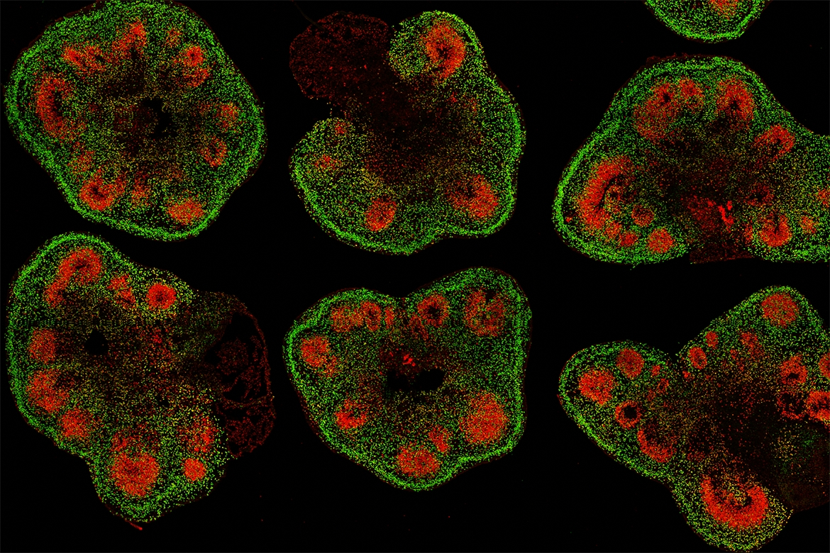
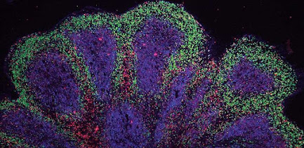
  • Defining the Architecture: Unlike traditional cell cultures, organoids are 3D structures grown from pluripotent stem cells (iPSCs) or adult stem cells. They rely on the cells' intrinsic ability to self-organize, creating complex structures that mimic the lineage and spatial arrangement of an in vivo organ.
  • The "Avatar" in the Lab: Organoids allow for Personalized Medicine. By growing an organoid from a specific patient's cells, researchers can test drug responses on a "digital twin" of that patient’s tumor or tissue, eliminating the guesswork of trial-and-error prescriptions.
  • Bridge to Clinical Trials: Organoids serve as a critical bridge between the Petri dish and human clinical trials, potentially reducing the failure rate of new drugs and decreasing the reliance on animal testing models which often fail to predict human reactions.
  • The Ethical Frontier: As cerebral organoids (mini-brains) become more complex, exhibiting brain waves similar to preterm infants, science faces a profound question: At what point does biological complexity become sentience?

Thursday, September 29, 2022

Making lab-grown brain organoids ‘brainier

 Slices of mini–brain organoids with neural stem cells (red) and cortical neurons (green).
Credit: Hajime Ozaki, Watanabe lab/UCI

By using stem cells to grow miniature brain-like organs in the lab, scientists have opened a new avenue for studies of neurological development, disease and therapies that can’t be conducted in living people. But not all mini–brain organoids are created equal and getting them to precisely mimic the human brain tissues they’re modeling has been a persistent challenge.

“Right now, it’s like the Wild West because there is no standard method for generating mini–brain organoids,” said Bennett Novitch, a member of the Eli and Edythe Broad Center of Regenerative Medicine and Stem Cell Research at UCLA and the senior author of a new paper on the topic. “Every neuroscientist wants to make a brain organoid model of their favorite disease, and yet everyone’s organoids do not always look alike.”

In fact, because there is no common protocol for their production and a lack of quality-control guidelines, organoids can vary from lab to lab — and even from batch to batch — which means that a finding made in one organoid may not hold true in another.

“If my lab and another lab down the hall were to conduct drug screens using mini–brain organoid models of the same disorder, we could still get different results,” said Momoko Watanabe, the new paper’s first author and an assistant professor of anatomy and neurobiology at UC Irvine. “We won’t know whose findings are correct because the differences we’re seeing could be reflections of how our models differ rather than reflections of the disease.”

Tuesday, May 16, 2023

Brain research with organoids

Section of an electroporated brain organoid of a common marmoset. Green: electroporated cells that glow green due to the green fluorescent protein; magenta: neurons; gray: nuclei.
Photo Credit: Lidiia Tynianskaia

Scientists at the German Primate Center develop effective method to genetically modify brain organoids

Primates are among the most intelligent creatures with distinct cognitive abilities. Their brains are relatively large in relation to their body stature and have a complex structure. However, how the brain has developed over the course of evolution and which genes are responsible for the high cognitive abilities is still largely unclear. The better our understanding of the role of genes in brain development, the more likely it will be that we will be able to develop treatments for serious brain diseases. 

Researchers are approaching these questions by knocking out or activating individual genes and thus drawing conclusions about their role in brain development. To avoid animal experiments as far as possible, brain organoids are used as an alternative. These three-dimensional cell structures, which are only a few millimeters in size, reflect different stages of brain development and can be genetically modified. However, such modifications are usually very complex, lengthy and costly. Researchers at the German Primate Center (DPZ) – Leibniz Institute for Primate Research in Göttingen have now succeeded in genetically manipulating brain organoids quickly and effectively. 

Thursday, April 7, 2022

Mini-Livers on a Chip

Researchers at Gladstone Institutes designed a new platform for studying how the human immune system responds to hepatitis C infection by combining microfluidic technology with liver organoids. Credit: Gladstone Institutes

A vaccine for hepatitis C has eluded scientists for more than 30 years, for several reasons. For one, the virus that causes the disease comes in many genetic forms, complicating the creation of a widely effective vaccine. For another, studying hepatitis C has been difficult because options in animals are limited and lab methods using infected cells have not adequately reflected the real-life dynamics of infection.

Now, researchers at Gladstone Institutes have developed a new platform for studying how the human immune system responds to hepatitis C infection. The method, presented in the scientific journal Open Biology, marries microfluidic technology (which allows scientists to precisely manipulate fluid at a microscopic scale) with liver organoids (three-dimensional cell clusters that mimic the biology of real human livers).

“The 3D structure and cellular composition of liver organoids allows us to study viral entry and replication in a highly relevant physiological manner,” says Gladstone Senior Investigator Todd McDevitt, PhD, a senior author of the new study.

“Our approach enables a more controlled and accurate investigation into the immune response to hepatitis C infection,” says Melanie Ott, MD, PhD, director of the Gladstone Institute of Virology and another senior author of the study. “We hope our method will accelerate the discovery of a much-needed vaccine.”

Tuesday, September 13, 2022

The gene to which we owe our big brain

A section of a brain organoid made from stem cells of a human. In magenta are actively proliferating brain stem cells, in yellow a subset of brain stem cells.
Photo Credit: Jan Fischer

ARHGAP11B - this complex name is given to a gene that is unique to humans and plays an essential role in the development of the neocortex. The neocortex is the part of the brain to which we owe our high mental abilities. A team of researchers from the German Primate Center (DPZ) - Leibniz Institute for Primate Research in Göttingen, the Max Planck Institute for Molecular Cell Biology and Genetics (MPI-CBG) in Dresden, and the Hector Institute for Translational Brain Research (HITBR) in Mannheim has investigated the importance of ARHGAP11B in neocortex development during human evolution. 

To do this, the team introduced for the first time a gene that exists only in humans into laboratory-grown brain organoids from our closest living relatives, chimpanzees. In the chimpanzee brain organoid, the ARHGAP11B gene led to an increase in brain stem cells relevant to brain growth and an increase in those neurons that play a critical role in the extraordinary mental abilities of humans. If, on the other hand, the ARHGAP11B gene was switched off in human brain organoids, the quantity of these brain stem cells fell to the level of a chimpanzee. Thus, the research team was able to show that the ARGHAP11B gene played a crucial role in the evolution of the brain from our ancestors to modern humans.

Tuesday, June 6, 2023

Fine-tuning 3D lab-grown mini tumors to help predict how patients respond to cancer therapies

The improved process allows researchers to use an advanced imaging method to study and analyze individual organoids in great detail.
Image Credit: Soragni Lab.

Scientists from the UCLA Jonsson Comprehensive Cancer Center have developed a new method to bio-print miniature tumor organoids that are designed to mimic the function and architecture of real tumors. The improved process allows researchers to use an advanced imaging method to study and analyze individual organoids in great detail, which can help researchers identify personalized treatments for people with rare or hard-to-treat cancers.

The method is described in the journal Nature Communications.

“Tumor organoids have become fundamental tools to investigate tumor biology and highlight drug sensitivities of individual patients,” said Alice Soragni, PhD, an assistant professor in the department of Orthopedic Surgery at the David Geffen School of Medicine at UCLA and member of the UCLA Jonsson Comprehensive Cancer Center. “However, we still need better ways to anticipate if resistance could be arising in a small population of cells, which we may not detect using conventional screening approaches. This is truly important, particularly as organoid-based drug predictions are starting to be leveraged clinically.”

Monday, September 13, 2021

Engineers grow pancreatic organoids

MIT and Cancer Research UK Manchester Institute researchers have
developed a synthetic gel that can be used to grow tiny
pancreatic organoids, seen here, from human pancreatic cells.
Credits: Courtesy of the researchers.

MIT engineers, in collaboration with scientists at Cancer Research UK Manchester Institute, have developed a new way to grow tiny replicas of the pancreas, using either healthy or cancerous pancreatic cells. Their new models could help researchers develop and test potential drugs for pancreatic cancer, which is currently one of the most difficult types of cancer to treat.

Using a specialized gel that mimics the extracellular environment surrounding the pancreas, the researchers were able to grow pancreatic “organoids,” allowing them to study the important interactions between pancreatic tumors and their environment. Unlike some of the gels now used to grow tissue, the new MIT gel is completely synthetic, easy to assemble and can be produced with a consistent composition every time.

Thursday, October 21, 2021

Lab-grown ‘mini brains’ hint at treatments for neurodegenerative diseases

Mini brain organoids showing cortical-like structures 
Credit: Andras Lakatos
A common form of motor neuron disease, amyotrophic lateral sclerosis, often overlaps with frontotemporal dementia (ALS/FTD) and can affect younger people, occurring mostly after the age of 40-45. These conditions cause devastating symptoms of muscle weakness with changes in memory, behavior and personality. Being able to grow small organ-like models (organoids) of the brain allows the researchers to understand what happens at the earliest stages of ALS/FTD, long before symptoms begin to emerge, and to screen for potential drugs.

In general, organoids, often referred to as ‘mini organs’, are being used increasingly to model human biology and disease. At the University of Cambridge alone, researchers use them to repair damaged livers, SARS-CoV-2 infection of the lungs and model the early stages of pregnancy, among many other areas of research.

Typically, researchers take cells from a patient’s skin and reprogram the cells back to their stem cell stage – a very early stage of development at which they have the potential to develop into most types of cell. These can then be grown in culture as 3D clusters that mimic particular elements of an organ. As many diseases are caused in part by defects in our DNA, this technique allows researchers to see how cellular changes – often associated with these genetic mutations – lead to disease.

Monday, May 8, 2023

A new at­las il­lus­trates how the hu­man ret­ina is de­vel­op­ing.

De­tail of a cross-​section of a ret­inal or­ganoid. Dif­fer­ent tis­sue struc­tures are made vis­ible with dif­fer­ent colors.
Pho­to­ Credit: Wahle et al. Nature Bi­o­tech­no­logy 2023

What cell types are found in which hu­man tis­sue, and where? Which genes are act­ive in the in­di­vidual cells, and which pro­teins are found there? An­swers to these ques­tions and more are to be provided by a specialized at­las – in par­tic­u­lar how the dif­fer­ent tis­sues form dur­ing em­bryonic de­vel­op­ment and what causes dis­eases. In cre­at­ing this at­las, re­search­ers aim to map not only tis­sue dir­ectly isol­ated from hu­mans, but also struc­tures called or­ganoids. These are three-​dimensional clumps of tis­sue that are cul­tiv­ated in the labor­at­ory and de­velop in a way sim­ilar to hu­man or­gans, but on a small scale.

“The ad­vant­age of or­ganoids is that we can in­ter­vene in their de­vel­op­ment and test act­ive sub­stances on them, which al­lows us to learn more about healthy tis­sue as well as dis­eases,” ex­plains Bar­bara Treut­lein, Pro­fessor of Quant­it­at­ive De­vel­op­mental Bio­logy at the De­part­ment of Biosys­tems Sci­ence and En­gin­eer­ing at ETH Zurich in Basel.

To help pro­duce such an at­las, Treut­lein, to­gether with re­search­ers from the Uni­ver­sit­ies of Zurich and Basel, has now de­veloped an ap­proach to gather and com­pile a great deal of in­form­a­tion about or­ganoids and their de­vel­op­ment. The re­search team ap­plied this ap­proach to the or­ganoids of the hu­man ret­ina, which they de­rived from stem cells.

Friday, March 29, 2024

Not unique to humans but uniquely human: researchers identify factor involved in brain expansion in humans

A microscopy image of a human brain organoid.
Image Credit: © Janine Hoffmann

What makes us human? According to neurobiologists it is our neocortex. This outer layer of the brain is rich in neurons and lets us do abstract thinking, create art, and speak complex languages. An international team led by Dr. Mareike Albert at the Center for Regenerative Therapies Dresden (CRTD) of TUD Dresden University of Technology has identified a new factor that might have contributed to neocortex expansion in humans. The results were published in the EMBO Journal.

The neocortex is the characteristic folded outer layer of the brain that resembles a walnut. It is responsible for higher cognitive functions such as abstract thinking, art, and language. “The neocortex is the most recently evolved part of the brain,” says Dr. Mareike Albert, research group leader at the CRTD. “All mammals have a neocortex, but it varies in size and complexity. Human and primate neocortices have folds while, for example, mice have a completely smooth neocortex, without any creases.”

The folds characteristic of the human brain increases the surface area of the neocortex. The human neocortex has a greater number of neurons that support complex cognitive functions.

The molecular mechanisms driving neocortex evolution are still largely unknown. “Which genes are responsible for inter-species differences in neocortex size? What factors have contributed to brain expansion in humans? Answering these questions is crucial to understanding human brain development and potentially addressing mental health disorders,” explains Dr. Albert.

Thursday, April 4, 2024

Scientists discover potential treatment approaches for polycystic kidney disease

cientists would like to know how cysts form in polycystic kidney disease (PKD). Here, they compared two 3-D mini-kidney models. On the left, a model shows a mini kidney with a gene mutation that causes cysts to form. On the right, researchers used gene editing to correct a gene mutation, preventing the development of cysts.
Image Credit: Vishy, et al., Cell Stem Cell 2024

Researchers have shown that dangerous cysts, which form over time in polycystic kidney disease (PKD), can be prevented by a single normal copy of a defective gene. This means the potential exists that scientists could one day tailor a gene therapy to treat the disease. They also discovered that a type of drug, known as a glycoside, can sidestep the effects of the defective gene in PKD. The discoveries could set the stage for new therapeutic approaches to treating PKD, which affects millions worldwide. The study, partially funded by the National Institutes of Health (NIH), is published in Cell Stem Cell.

Scientists used gene editing and 3-D human cell models known as organoids to study the genetics of PKD, which is a life-threatening, inherited kidney disorder in which a gene defect causes microscopic tubes in the kidneys to expand like water balloons, forming cysts over decades. The cysts can crowd out healthy tissue, leading to kidney function problems and kidney failure. Most people with PKD are born with one healthy gene copy and one defective gene copy in their cells.

“Human PKD has been so difficult to study because cysts take years and decades to form,” said senior study author Benjamin Freedman, Ph.D., at the University of Washington, Seattle. “This new platform finally gives us a model to study the genetics of the disease and hopefully start to provide answers to the millions affected by this disease.”

Tuesday, November 2, 2021

‘Live’ brain models used in hunt for Alzheimer’s treatment

Alzheimer plaques in human stem cell derived neurons
Studying tiny ‘live’ models of the human brain has helped researchers understand its ageing and find a key to potential treatments for Alzheimer’s and other neurodegenerative diseases.

University of Queensland scientists have found different cellular mechanisms that can either accelerate or reduce brain cell deterioration.

Professor Ernst Wolvetang studied organoids, models that closely mimic the human brain, at UQ’s Australian Institute for Bioengineering and Nanotechnology.

“We have found that human brain organoids can be used to study the molecular mechanisms that drive brain ageing processes,” Professor Wolvetang said.

“This opens the way for testing many molecules that could become potential therapeutic drugs for a host of neurodegenerative diseases.”

Using the organoids, Professor Wolvetang and Dr Julio Aguado found DNA leakage accelerated ageing in the rare neurodegenerative disease Ataxia-Telangiectasia (A-T).

In another research project, Professor Wolvetang and Dr Mohammed Shaker found that increasing levels of the ‘anti-ageing’ protein klotho reduced the deterioration in brain cells associated with age and dementia.

Wednesday, October 15, 2025

Did Lead Limit Brain and Language Development in Neanderthals and Other Extinct Hominids?

UC San Diego researchers have found high levels of lead in the teeth of both Neanderthals (left) and modern humans (right). However, a gene mutation may have protected modern human brains, allowing language to flourish.
Photo Credit: Kyle Dykes/UC San Diego Health Sciences

Ancient human relatives were exposed to lead up to two million years ago, according to a new study. However, a gene mutation may have protected modern human brains, allowing language to flourish.

What set the modern human brain apart from our now extinct relatives like Neanderthals? A new study by University of California San Diego School of Medicine and an international team of researchers reveals that ancient hominids — including early humans and great apes — were exposed to lead earlier than previously thought, up to two million years before modern humans began mining the metal. This exposure may have shaped the evolution of hominid brains, limiting language and social development in all but modern humans due to a protective genetic variant that only we carry. The study was published in Science Advances.

The researchers analyzed fossilized teeth from 51 hominids across Africa, Asia and Europe, including modern and archaic humans such as Neanderthals, ancient human ancestors like Australopithecus africanus, and extinct great apes such as Gigantopithecus blacki.

Thursday, December 25, 2025

Restoring the healthy form of a protein could revive blood vessel growth in premature infants’ lungs

A blood vessel organoid.
Video Credit: Yunpei Zhang and Enbo Zhu, Mingxia Gu Lab

A UCLA-led research team has discovered a molecular switch that determines whether tiny blood vessels in premature infants’ lungs can regenerate after injury. A failure of this repair process is a hallmark of bronchopulmonary dysplasia, or BPD, a serious lung disease that affects babies born very early. It arises from a combination of premature birth, inflammation or infection, and exposure to the high levels of oxygen and breathing support that are necessary to keep these infants alive during a critical period of lung development.

The researchers found that in BPD, the blood vessel cells in the lungs begin producing a shortened, nonfunctional isoform — a version of a protein — called NTRK2, which has been extensively studied in the nervous system but not in the pulmonary vasculature. When this shortened isoform dominates, the lung cannot rebuild the delicate network of tiny blood vessels needed for healthy breathing.

Monday, December 15, 2025

Reproduced human neural circuits show the crucial role of the thalamus in shaping the cortical circuit

Assembloid [3D fluorescent staining] Axons in the thalamus (pink) extended toward the cortex, while those in the cortex (green) extended toward the thalamus at 14 days post-fusion.
Image Credit: Fumitaka Osakada

A Japanese research team has successfully reproduced the human neural circuit in vitro using multi-region miniature organs known as assembloids, which are derived from induced pluripotent stem (iPS) cells. With this circuit, the team demonstrated that the thalamus plays a crucial role in shaping cell type-specific neural circuits in the human cerebral cortex.

These findings were published in the journal Proceedings of the National Academy of Sciences of the United States of America.

Our brain’s cerebral cortex contains various types of neurons, and effective communication among these neurons and other brain regions is crucial for activating functions like perception and cognition.

Patients with neurodevelopmental disorders, such as autism spectrum disorder (ASD), exhibit disruptions in the structure and function of neural circuits in the cerebral cortex. Therefore, understanding the principles of these circuits is essential to uncovering the causes of these disorders and developing new medications.

Wednesday, December 13, 2023

New treatment for deadly uterine cancer

left to right, Dr Asmerom Sengal, Professor Pamela Pollock.
Photo Credit: Courtesy of Queensland University of Technology

QUT scientists have discovered a promising new therapy for a deadly type of endometrial cancer that has a poor prognosis if the cancer spreads or returns after initial treatment, a plight that affects 15-20 per cent of endometrial cancer patients.

  • Testing of new drug inhibited uterine tumor cell growth in lab and mice models
  • The drug blocks the receptor of the growth factor in tumors that is associated with a low survival rate
  • The inhibitor also reduced the tumors blood vessel formation

Dr Asmerom Sengal and Associate Professor Pamela Pollock from QUT’s School of Biomedical Sciences, published their research in Nature Precision Oncology with a recommendation that the strength of their findings indicated they should proceed to patient trials.

Dr Asmerom said endometrial cancer confined within the uterus could be cured with surgery however, if it had spread to the abdomen and other organs patients had limited treatment options.

“Previously, we found women with endometrial cancer who have an incorrect growth factor receptor called fibroblast growth factor receptor 2c (FGFR2c) on the tumor cell surface have a poor survival rate,” Dr Asmerom said.

Thursday, November 13, 2025

A cellular protein, FGD3, boosts breast cancer chemotherapy, immunotherapy

The research team included, front row, from left: graduate student Junyao Zhu, biochemistry professor David Shapiro, and senior researcher Chengiian Mao; back row, from left: graduate students Abigail Spaulding, Xinyi Dai and Qianjin Jiang.
Photo Credit: Fred Zwicky

A naturally occurring protein that tends to be expressed at higher levels in breast cancer cells boosts the effectiveness of some anticancer agents, including doxorubicin, one of the most widely used chemotherapies, and a preclinical drug known as ErSO, researchers report. The protein, FGD3, contributes to the rupture of cancer cells disrupted by these drugs, boosting their effectiveness and enhancing anticancer immunotherapies.

The new findings were the happy result of experiments involving ErSO, an experimental drug that killed 95-100% of estrogen-receptor-positive breast cancer cells in a mouse model of the disease. ErSO upregulates a cellular pathway that normally protects cancer cells from stress, said University of Illinois Urbana-Champaign biochemistry professor David Shapiro, who led the new work with Illinois graduate student Junyao Zhu. But when that protective pathway is ramped up, the system goes awry.

Thursday, December 25, 2025

The Quest for the Synthetic Synapse

Spike Timing" difference (Biology vs. Silicon)
Image Credit: Scientific Frontline

The modern AI revolution is built on a paradox: it is incredibly smart, but thermodynamically reckless. A large language model requires megawatts of power to function, whereas the human brain—which allows you to drive a car, debate philosophy, and regulate a heartbeat simultaneously—runs on roughly 20 watts, the equivalent of a dim lightbulb.

To close this gap, science is moving away from the "Von Neumann" architecture (where memory and processing are separate) toward Neuromorphic Computing—chips that mimic the physical structure of the brain. This report analyzes how close we are to building a "synthetic synapse."

Tuesday, March 21, 2023

UCLA-led study uses base editing to correct mutation that causes rare immune deficiency

Image Credit: Sangharsh Lohakare

A new UCLA-led study suggests that advanced genome editing technology could be used as a one-time treatment for the rare and deadly genetic disease CD3 delta severe combined immunodeficiency.

The condition, also known as CD3 delta SCID, is caused by a mutation in the CD3D gene, which prevents the production of the CD3 delta protein that is needed for the normal development of T cells from blood stem cells.

Without T cells, babies born with CD3 delta SCID are unable to fight off infections and, if untreated, often die within the first two years of life. Currently, bone marrow transplant is the only available treatment, but the procedure carries significant risks.

In a study published in Cell, the researchers showed that a new genome editing technique called base editing can correct the mutation that causes CD3 delta SCID in blood stem cells and restore their ability to produce T cells.

The potential therapy is the result of a collaboration between the laboratories of Dr. Donald Kohn and Dr. Gay Crooks, both members of the Eli and Edythe Broad Center of Regenerative Medicine and Stem Cell Research at UCLA and senior authors of the study.

Monday, February 26, 2024

Human stem cells coaxed to mimic the very early central nervous system

Jianping Fu, Ph.D., Professor of Mechanical Engineering at the University of Michigan and the corresponding author of the paper being published at Nature discusses his team’s work in their lab with Jeyoon Bok, Ph.D. candidate at the Department of Mechanical Engineering.
Photo Credit: Marcin Szczepanski, Michigan Engineering

The first stem cell culture method that produces a full model of the early stages of the human central nervous system has been developed by a team of engineers and biologists at the University of Michigan, the Weizmann Institute of Science, and the University of Pennsylvania.

“Models like this will open doors for fundamental research to understand early development of the human central nervous system and how it could go wrong in different disorders,” said Jianping Fu, U-M professor of mechanical engineering and corresponding author of the study in Nature.

The system is an example of a 3D human organoid—stem cell cultures that reflect key structural and functional properties of human organ systems but are partial or otherwise imperfect copies.

“We try to understand not only the basic biology of human brain development, but also diseases—why we have brain-related diseases, their pathology, and how we can come up with effective strategies to treat them,” said Guo-Li Ming, who along with Hongjun Song, both Perelman Professors of Neuroscience at UPenn and co-authors of the study, developed protocols for growing and guiding the cells and characterized the structural and cellular characteristics of the model.

Featured Article

Hidden heartache of losing an animal companion

Chimmi April 09, 2010 -February 23, 2025 My best friend. Photo Credit: Heidi-Ann Fourkiller The emotional toll of losing a beloved pet durin...

Top Viewed Articles