. Scientific Frontline: Neuroscience
Showing posts with label Neuroscience. Show all posts

Wednesday, August 31, 2022

3D imaging contributes to a better understanding of early stages of Alzheimer's disease

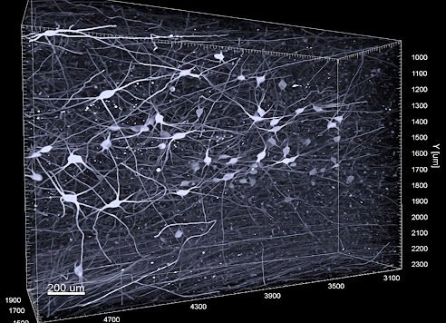
Three-dimensional image of noradrenergic nerve cells in the envelope of locus coeruleus.
Photo credit: Gilvesy et al.

With the help of a new imaging technique for 3D, researchers at Karolinska Institutet, among others, have been able to characterize a part of the brain that shows the most accumulation of tau protein, an important biomarker for the development of Alzheimer's disease. The results published in the journal Acta Neuropathologica may in the future make it possible to have a more accurate neuropathological diagnosis of Alzheimer's disease spectrum at a very early stage.

Intracellular accumulation of pathological tau protein in the brain is a hallmark of several age-related neurodegenerative diseases, including Alzheimer's disease, which accounts for 60-80 percent of all dementia cases worldwide.

In a new study, researchers at Karolinska Institutet, SciLifeLab in Stockholm and several universities from Hungary, Canada, Germany and France have applied a state-of-the-art immune imaging technology, in combination with light sheet microscopy, to investigate a human brain stem core, locus coeruleus, which is an important core in the mammalian brain.

Tuesday, August 23, 2022

Sleepless and selfish: Lack of sleep makes us less generous

The new study shows how sleep loss dramatically reduces the desire to help others, triggered by a breakdown in the activity of key prosocial brain networks.
Image credit: Eti Ben Simon and Matthew Walker, UC Berkeley

Humans help each other — it’s one of the foundations of civilized society. But a new study by scientists at the University of California, Berkeley, reveals that a lack of sleep blunts this fundamental human attribute, with real-world consequences.

Lack of sleep is known to be associated with an increased risk of cardiovascular disease, depression, diabetes, hypertension and overall mortality. However, these new discoveries show that a lack of sleep also impairs our basic social conscience, making us withdraw our desire and willingness to help other people.

In one portion of the new study, the scientists showed that charitable giving in the week after the beginning of Daylight-Saving Time, when residents of most states “spring forward” and lose one hour of their day, dropped by 10% — a decrease not seen in states that do not change their clocks or when states return to standard time in the fall.

The study, led by UC Berkeley research scientist Eti Ben Simon and Matthew Walker, a UC Berkeley professor of psychology, adds to a growing body of evidence demonstrating that inadequate sleep not only harms the mental and physical well-being of an individual, but also compromises the bonds between individuals — and even the altruistic sentiment of an entire nation.

Wednesday, August 17, 2022

Researchers reprogram human skin cells to aged neurons to study neurodegenerative disorders

CC0 Public Domain

Researchers at Lund University in Sweden have developed a new method for studying age-related brain disorders. The researchers have focused on the neurodegenerative disorder Huntington’s disease and the results have now been published in the journal Brain.

Basic medical research often faces the challenge of developing disease models that correspond to specific disease mechanisms or the disease to be studied. This is a challenge that needs to be solved in order to produce new effective treatments. One example of a disease that is difficult to model for an understanding of the underlying mechanisms is Huntington’s disease. In part, this is due to the difficulty in recreating adequate animal or cellular models.

By reprogramming skin cells into neurons, Johan Jakobsson and his research group have been able to study Huntington’s disease in an innovative way that he believes could be significant for successful studies of several age-related brain disorders.

“We took skin biopsies from patients living with Huntington’s disease and reprogrammed the skin biopsies into neurons. We then compared these neurons with reprogrammed neurons from healthy people. The results are very interesting. We have found several defects that explain some of the disease mechanisms in neurons from patients with Huntington’s disease. Among other things, we observed that neurons from patients with Huntington’s disease show problems in breaking down and recycling a particular kind of protein – which can lead to a lack of energy in these cells”, says Johan Jakobsson, professor of neuroscience at Lund University.

Wednesday, August 10, 2022

Ageing neutralizes sex differences in the brain

When male and female fruit flies age, their brains become desexualized.
Credit: Erik Karits on Unsplash

When male and female fruit flies age, their brains become desexualized. Age-related changes take place in both sexes, but the male brain becomes feminized to a larger extent than the female brain becomes masculinized. This is the conclusion of a study performed by a research group at Linköping University.

It is a well-known fact that weaker individuals cannot afford to “invest” in sexual behaviors to the same extent as their healthier conspecifics. However, it is not clear if ageing, which weakens individuals, also leads to a reduced investment in sexual activities. You might think that for individuals close to the end of their lives, going “all in” on reproduction, in order to pass on their genes before it is too late, would be best. Sexual behaviors are directed from the brain, and to find out what happens to sex differences in this tissue when fruit flies age the researchers have investigated how genes expressed to different degrees in young males and females change over time.

“Our results show that gene expression in male and female brains become more similar with age, and that both sexes contribute to this pattern”, says Dr Antonino Malacrinò, one of the study’s main authors who now works at the University of Reggio Calabria in Italy.

What the study shows is that if the expression of a certain gene is higher in the brains of young females than in young males, the gene’s expressions is reduced in older females and increased in old males – and vice versa for genes with higher expression in young males.

“The results also show that the changes are larger in males than in females”, says Antonino Malacrinò.

Secret behind ‘nic-sickness’ could help break tobacco addiction

Nicotine is addictive because it activates the brain’s dopamine network, which makes us feel good. UC Berkeley researchers now show in experiments on mice that nicotine in high doses also activates a recently discovered dopamine network that responds to unpleasant stimuli. This aversive dopamine network could be leveraged to create a therapy that boosts the negative effects and lessens the rewards of nicotine.
Image credit: Christine Liu, UC Berkeley

If you remember your first hit on a cigarette, you know how sickening nicotine can be. Yet, for many people, the rewards of nicotine outweigh the negative effects of high doses.

University of California, Berkeley, researchers have now mapped out part of the brain network responsible for the negative consequences of nicotine, opening the door to interventions that could boost the aversive effects to help people quit smoking.

Though most addictive drugs at high doses can cause physiological symptoms that lead to unconsciousness or even death, nicotine is unique in making people physically ill when inhaled or ingested in large quantities. As a result, nicotine overdoses are rare, though the advent of e-cigarettes has made “nic-sick” symptoms like nausea and vomiting, dizziness, rapid heartbeat and headaches more common.

New research, conducted in mice, suggests that this aversive network could be manipulated to treat nicotine dependence.

“Decades of research have focused on understanding how nicotine reward leads to drug addiction and what are the underlying brain circuits. In contrast, the brain circuits that mediate the aversive effects of nicotine are largely understudied,” said Stephan Lammel, UC Berkeley associate professor of molecular and cell biology. “What we found is that the brain circuits that are activated after a high aversive dose are actually different from those that are activated when nicotine is delivered at a low dose. Now that we have an understanding of the different brain circuits, we think we can maybe develop a drug so that, when nicotine is taken at a low dose, these brain circuits can be coactivated to induce an acute aversive effect. This could actually be a very effective treatment for nicotine addiction in the future, which we currently do not have.”

Tuesday, August 9, 2022

A role for cell ‘antennae’ in managing dopamine signals in the brain

Microscope image of a cultured mouse neuron from the striatum region of the brain labeled with a green fluorescent antibody that detects dopamine receptor 1. The receptor localizes along the cell surface and is enriched in a primary cilium projecting from the cell body. Nuclei are indicated in blue.
 Credit: Image courtesy of Kirk Mykytyn

A historically overlooked rod-like projection present on nearly every cell type in the human body may finally be getting its scientific due: A new study has found that these appendages, called cilia, on neurons in the brain have a key role in ensuring a specific dopamine receptor’s signals are properly received.

The research was conducted in mouse models of a disorder called Bardet-Biedl syndrome, and applies to one of five proteins that regulate dopamine signaling, called dopamine receptor 1. In certain regions of the brain, this receptor can be thought of as an “on” switch that initiates motivated behavior – basically any behavior linked to pursuit of a goal.

The study showed that if the receptor either gets stuck on cilia or never has a chance to localize to these cell “antennae,” messages telling the body to move are reduced.

“There’s something about dopamine receptor 1 needing to get to and from neuronal cilia that’s required for proper signaling,” said lead author Kirk Mykytyn, associate professor of biological chemistry and pharmacology in The Ohio State University College of Medicine. “This is the first demonstration that cilia are important for dopamine receptor 1 signaling.”

Sunday, August 7, 2022

When a task adds more steps, this circuit helps you notice

In their study, researchers traced neurons projecting from the anterior cingulate cortex (right, red) to the motor cortex (left, green). Note the images are at different scales.
Source: Picower Institute for Learning and Memory

Life is full of processes to learn and then relearn when they become more elaborate. One day you log in to an app with just a password, then the next day you also need a code texted to you. One day you can just pop your favorite microwavable lunch into the oven for six straight minutes, but then the packaging changes and you have to cook it for three minutes, stir, and then heat it for three more. Our brains need a way to keep up. A new study by neuroscientists at The Picower Institute for Learning and Memory at MIT reveals some of the circuitry that helps a mammalian brain learn to add steps.

In Nature Communications the scientists report that when they changed the rules of a task, requiring rats to adjust from performing just one step to performing two, a pair of regions on the brain’s surface, or cortex, collaborated to update that understanding and change the rats’ behavior to fit the new regime. The anterior cingulate cortex (ACC) appeared to recognize when the rats weren’t doing enough and updated cells in the motor cortex (M2) to adjust the task behavior.

“I started this project about 7 or 8 years ago when I wanted to study decision making.” said Daigo Takeuchi, a researcher at the University of Tokyo who led the work as a postdoc at the RIKEN-MIT Laboratory for Neural Circuit Genetics at The Picower Institute directed by senior author and Picower Professor Susumu Tonegawa. “New studies were finding a role for M2. I wanted to study what upstream circuits were influencing this.”

How bat brains listen out for incoming signals during echolocation

Bats "see" with the ears. Scientists at Goethe University have found out how the auditory cortex is prepared for the incoming acoustic signals.
Credit: Hechavarria

When bats emit sounds for echolocation, a feedback loop modulates the sensitivity of the auditory cortex for incoming acoustic signals. Neuroscientists from the Goethe University Frankfurt found out. In a study published in the journal "Nature Communications", they show that the flow of information in the neuronal circuit involved reversed as the sound was generated. This feedback prepares the auditory cortex for the expected “echoes” of the sounds sent out. The researchers see their results as a sign that the importance of feedback loops in the brain is currently still underestimated.

Bats are famous for their ultrasound navigation: they orientate themselves through their extremely sensitive hearing by emitting ultrasound sounds and getting a picture of their environment based on the sound thrown back. For example, the eyelid nose bat (Carollia perspicillata) the fruits she prefers as food through this echolocation system. At the same time, the bats also use their voice to communicate with their peers, for which they choose a somewhat lower frequency range.

Neuroscientist Julio C. Hechavarria from the Institute for Cell Biology and Neuroscience at Goethe University, together with his team, examines which brain activities in the case of the eyewear nose go hand in hand with the vocalizations. In their latest study, the Frankfurters examined how the front lobes - a region in the front brain that is associated with the planning of actions in humans - and the auditory cortex, in which acoustic signals are processed, work together in the echolocation. For this purpose, the researchers used tiny electrodes on the bats, which recorded the activity of the nerve cells in the frontal lobe and in the auditory cortex.

Wednesday, June 22, 2022

Brain imaging links stimulant-use relapse to distinct nerve pathway

Researchers used advanced brain imaging techniques to study nerve fibers connecting to the nucleus accumbens, which plays an important role in motivation and addiction.
Credit: Loreen Tisdall and Kelly H. MacNiven.

You might assume that people who are most prone to developing a substance use disorder in the first place would also have the hardest time avoiding relapse following treatment. But a new study by scientists with the Wu Tsai Neurosciences Institute’s NeuroChoice Initiative reveals that relapse may be linked to quite different brain circuits than addiction itself.

“There’s a huge revolving door problem with relapse,” said Brian Knutson, a professor of psychology. “These findings suggest ​​that what gets you into taking drugs may not be the same processes that get you out of it, which could be very valuable to help predict who is at highest risk of relapse coming out of treatment.”

Drug addiction presents a major global challenge. More than 35 million people worldwide self-report problematic use of drugs and admissions to drug treatment programs have surged in the United States in recent years. For many drugs, in particular stimulants such as cocaine and amphetamines, relapse remains a common problem. For example, as many as 50 percent of people with stimulant use disorders relapse within 6 months of release from treatment.

“The statistics are disheartening,” said Kelly MacNiven, a social science research scholar in the Knutson lab and co-author of the new study. “Unfortunately not much is known at a biological level about the drivers of relapse — understanding this better is going to be the first step to developing better ways to help people get out of dependence.”

Monday, June 20, 2022

Gene discovery indicates motor neuron diseases caused by abnormal lipid processing in cells

A new genetic discovery adds weight to a theory that motor neuron degenerative diseases are caused by abnormal lipid (fat) processing pathways inside brain cells. This theory will help pave the way for new diagnostic approaches and treatments for this group of conditions. The discovery will provide answers for certain families who have previously had no diagnosis.

Motor neuron degenerative diseases (MNDs) are a large family of neurological disorders. Currently, there are no treatments available to prevent onset or progression of the condition. MNDs are caused by changes in one of numerous different genes. Despite the number of genes known to cause MNDs, many patients remain without a much-needed genetic diagnosis.

The team behind the current work developed a hypothesis to explain a common cause of MNDs stemming from their discovery of 15 genes responsible for MNDs. The genes they identified are all involved in processing lipids - in particular cholesterol – inside brain cells. Their new hypothesis, published in the journal Brain, describes the specific lipid pathways that the team believe are important in the development of MNDs.

Now, the team has identified a further new gene – named TMEM63C – which causes a degenerative disease that affects the upper motor neuron cells in the nervous system. Also published in Brain, their latest discovery is important as the protein encoded by TMEM63C is located in the region of the cell where the lipid processing pathways they identified operate. This further bolster the hypothesis that MNDs are caused by abnormal processing of lipids including cholesterol.

“This new gene finding is consistent with our hypothesis that the correct maintenance of specific lipid processing pathways is crucial for the way brain cells function, and that abnormalities in these pathways are a common linking theme in motor neuron degenerative diseases,” said study co-author Professor Andrew Crosby from the University of Exeter. “It also enables new diagnoses and answers to be readily provided for families affected by some forms of MND”

New imaging technique to find out what happens in the brains of dogs and cats

In a preliminary experiment, Parkkonen held a quantum optical MEG sensor with his hand on his family cat’s, Roosa’s, head while she listened to simple sound sequences.
Credit: Professor Lauri Parkkonen / Aalto University

For years, Professor Lauri Parkkonen's team at Aalto University has been developing quantum optical sensors for measuring the brain's magnetic fields using a technique known as magnetoencephalography (MEG). In traditional MEG, the superconducting sensors operate at very low temperatures and need centimeters of thermal insulation, but the quantum optical sensors work at room temperature, so they can be placed directly on the surface of the head. This allows more accurate measurements of the brain’s magnetic fields.

Parkkonen and his team plan to use the new method to build on their earlier work measuring brain activity in cats and dogs. Now they plan to characterize the complexity of the temporal structures in sensory stimuli that cat and dog brains can track. Similar experiments in humans have found that our brain produces specific responses to deviations in complex structures only when we attend to the stimuli and become aware of the deviations. Once the technique is perfected, Parkkonen and his team plan to use it to make similar measurements in human babies.

The experiments will begin this autumn – though Parkkonen has already done some preliminary tests with his family cat, Roosa – and the project is expected to continue until 2026. The researchers hope that their findings will provide an unprecedented window onto the cognition of cats and dogs, and this could also help bridge the gap between our understanding of human brains and the brains of other mammals.

Friday, June 17, 2022

Tenascin proteins inhibit cell sheath regeneration

Juliane Bauch (left) and Andreas Faissner from the Chair of Cell Morphology and Molecular Neurobiology
Credit: RUB, Kramer

In multiple sclerosis, nerve cells lose their insulating layer. Researchers from Bochum are looking for starting points to promote regeneration processes. They have identified two relevant proteins.

Researchers at the Ruhr University Bochum have investigated the role that the two proteins tenascin C and tenascin R play in multiple sclerosis. In the disease, cells of the immune system destroy the myelin sheaths, i.e. the sheathing of the nerve cells. As the Bochum team showed in experiments with mice, the presence of the two Tenascins inhibits the regeneration of the myelin sheaths. Dr. Juliane Bauch and Prof. Dr. Andreas Faissner from the Bochum Chair for Cell Morphology and Molecular Neurobiology describes the results in the journal Cells.

The cause of the destruction of myelin sheaths in multiple sclerosis has not yet been clarified. "But the organism has various mechanisms to partially compensate for the lesions," says Juliane Bauch, who dealt intensively with the topic in her doctorate. The aim of the work is to identify starting points with which the regeneration of myelin sheaths could be improved.

Thursday, June 16, 2022

Parasitic worms reveal new insights into the evolution of sex and sex chromosomes

Schematic picture designed by the authors: During evolution, different chromosome elements represented by the Lego bricks (NA, NB, NX..etc. in the figure) were added onto the ancestral sex chromosomes in different species, forming the great diversity of nematode sex chromosomes. These "Lego" combinations of chromosome elements are shown as corresponding colors for each ‘clade’ of nematode species.
Credit: Quzijian

Studying two highly divergent phyla of worms that contain numerous parasites that cause human and livestock diseases, the research group of Qi Zhou from the University of Vienna and Zhejiang University, sheds light on how sexual reproduction and subsequent great diversity of sex chromosomes might have evolved.

Animals or plants with separate sexes are widespread in nature, and result from independent transitions from their hermaphroditic ancestor. The actual mechanism involved in the transitions between asexual and sexual reproductive modes, in other words, how is sex originated, remains an important and unanswered question. Excluding insects, about one third of the animal species, such as earthworms, snails, and some teleosts, are hermaphroditic. A comparison with their relatives who have evolved separate sexes, might reveal, how this particular trait originated and evolved in animals.

A new paper in Nature Communications, published by Qi Zhou of Department of Neuroscience and Developmental Biology in University of Vienna and Zhejiang University in China, provides clues into how separate sexes originated and characterizes how sex chromosomes evolved in flatworms or roundworms.

Tuesday, June 14, 2022

AI detects autism speech patterns across different languages

The researchers believe their work could provide a tool that might one day transcend cultures, because of the computer’s ability to analyze words and sounds in a quantitative way regardless of language.
 Photo Credit: by Emily Wade on Unsplash

A new study led by Northwestern University researchers used machine learning — a branch of artificial intelligence — to identify speech patterns in children with autism that were consistent between English and Cantonese, suggesting that features of speech might be a useful tool for diagnosing the condition.

Undertaken with collaborators in Hong Kong, the study yielded insights that could help scientists distinguish between genetic and environmental factors shaping the communication abilities of people with autism, potentially helping them learn more about the origin of the condition and develop new therapies.

Children with autism often talk more slowly than typically developing children, and exhibit other differences in pitch, intonation and rhythm. But those differences (called “prosodic differences'' by researchers) have been surprisingly difficult to characterize in a consistent, objective way, and their origins have remained unclear for decades.

However, a team of researchers led by Northwestern scientists Molly Losh and Joseph C.Y. Lau, along with Hong Kong-based collaborator Patrick Wong and his team, successfully used supervised machine learning to identify speech differences associated with autism.

Monday, June 13, 2022

Brain hereditary disease factor suspected

Jonasz Jeremiasz Weber, Rana Dilara Incebacak Eltemur, Priscila Pereira Sena, Huu Phuc Nguyen (from left) worked out the study together.
Credit: © Pengfei Qi

Similar to Alzheimer's, the hereditary disease Spinocerebellar Ataxia Type 17 (SCA17) leads to the demise of brain nerve cells and the premature death of those affected. The exact mechanisms of the disease are unknown, so there are no treatment approaches to date. Researchers of human genetics at the Ruhr University Bochum (RUB) around Dr. Jonasz Weber now suspects a class of protein-splitting enzymes, so-called calpaines, to contribute to the disease. In the model, the Calpaine was switched off to stop the course. The researchers report in the journal Cellular and Molecular Life Sciences.

Changed blueprint of a protein

Spinocerebellar ataxia type 17 (SCA17) is a rare, hereditary disease of the human brain. Due to the pathological change in a gene that contains the blueprint for a protein called TATA box-binding protein (TBP), the protein is formed in cells in a defective form. This also affects its function. "One consequence of this is that the protein forms detectable protein deposits in the brain and damages the nerve cells via molecular mechanisms that have not yet been fully elucidated," explains Jonasz Weber.

As a consequence, those affected by the disease develop symptoms such as movement disorders, seizures, impairment of mental performance as well as changes in nature and behavior, which are associated with the breakdown of tissues such as the cerebellum and brain stem.

Sunday, June 12, 2022

IA leads the charge against multiple sclerosis

MRI image in false colors of a brain hemisphere from an MS patient (affected areas are shown in red).
 Credit: Govind Bhagavatheeshwaran, Daniel Reich / NINDS / NIH

Artificial intelligence may enable earlier diagnosis of Multiple Sclerosis, an incurable disease that attacks the central nervous system. This could improve the efficacy of treatments designed to slow its progression.

An autoimmune disease, multiple sclerosis (MS) is characterized by a breakdown of myelin, the membrane that protects the axons of neurons. Communication within the nervous system is gradually disrupted, causing increasingly severe motor and neurological damage. Although multiple sclerosis is currently incurable, treatments are available to relieve certain symptoms, particularly if the disease is discovered early; unfortunately, however, it tends to be diagnosed at a later stage.

Scientists Have Found Neurons that Control Some Symptoms of Sickness

During an infection, inflammatory signals activate immune-sensitive neurons (genetically labeled in red) in the ventral medial preoptic area (VMPO) leading to the induction of fever and other sickness behaviors. All cells are labeled with a nuclear stain (blue).
Credit: Courtesy of Dulac Lab/HHMI at Harvard University

Feeling ill is about both the body and the brain. Now scientists have identified a group of neurons in mice that has ultimate control over symptoms such as fever and behaviors like seeking out warmth.

Fevers, chills, an appetite that vanishes – we can tell when we’re getting sick. Many people chalk these symptoms of illness up to the immune system fighting off infection. But there’s another player involved when we feel woefully under the weather.

“All of this is orchestrated by the brain,” says neurobiologist Catherine Dulac, who is a Howard Hughes Medical Institute Investigator at Harvard University. Now research from Dulac’s team, published in Nature, pins this broad response on a previously uncharacterized population of neurons in the brain.

How exactly the brain serves as an infection ringleader has been unclear. Earlier research had identified receptors in the brain that were required for animals to develop a fever. But fever is only part of the story. One of the bigger mysteries is: Where does ultimate control for the symptoms and behaviors associated with sickness lie?

Dulac, her postdoctoral fellow, Jessica A. Osterhout, and colleagues injected mice with molecules that mimic bacterial or viral infections to investigate that question. As the mice’s immune systems reacted to these inflammatory molecules, the researchers homed in on which neurons jumped into action. The team watched neurons’ gene expression through single-cell RNA sequencing and mapped the whereabouts of those neurons using a visualization technique called MERFISH, which was developed in the lab of HHMI Investigator Xiaowei Zhuang at Harvard, a collaborator in this work.

Thursday, June 9, 2022

Social isolation may impact brain volume in regions linked to higher risk of dementia

Elderly woman in the middle stages of Alzheimer 
Credit: Steven HWG

Social isolation is linked to lower brain volume in areas related to cognition and a higher risk of dementia, according to research published today in Neurology. The study found that social isolation was linked to a 26% increased risk of dementia, separately from risk factors like depression and loneliness.

“Social isolation is a serious yet underrecognized public health problem that is often associated with old age,” said study author Professor Jianfeng Feng of Fudan University in Shanghai, China. “In the context of the COVID-19 pandemic, social isolation, or the state of being cut off from social networks, has intensified. It’s more important than ever to identify people who are socially isolated and provide resources to help them make connections in their community.”

The study looked at over 460,000 people across the United Kingdom with an average age of 57 at the beginning of the study who were followed for nearly 12 years before the pandemic. Of those, almost 42,000 (9%) reported being socially isolated, and 29,000 (6%) felt lonely. During the study, almost 5,000 developed dementia.

Researchers collected survey data from participants, along with a variety of physical and biological measurements, including MRI data. Participants also took thinking and memory tests to assess their cognitive function. For social isolation, people were asked three questions about social contact: whether they lived with others; whether they had visits with friends or family at least once a month; and whether they participated in social activities such as clubs, meetings or volunteer work at least once a week. People were considered socially isolated if they answered no to at least two questions.

Visual system brain development implicated in infants who develop autism

Anatomical locations of the splenium (yellow) and right middle occipital gyrus (red) in a representative infant brain.
Source: University of North Carolina at Chapel Hill

For the first time, scientists have found that brain differences in the visual brain systems of infants who later develop autism are associated with inherited genetic factors.

Published in the American Journal of Psychiatry, this research shows that brain changes in the size, white matter integrity and functional connectivity of the visual processing systems of six-month-olds are evident well before they show symptoms of autism as toddlers. Moreover, the presence of brain changes in the visual system is associated with the severity of autism traits in their older siblings.

Led by Dr. Jessica Girault, assistant professor of psychiatry at the UNC School of Medicine, this is the first research to observe that infants with older siblings who have autism and who themselves later develop autism as toddlers have specific biological differences in visual processing regions of the brain and that these brain characteristics precede the appearance of autistic symptoms. The presence of those visual processing differences is related to how pronounced the autism traits are in the older siblings.

“We’re beginning to parse differences in infant brain development that might be related to genetic factors,” said Girault, who is also a member of the Carolina Institute of Developmental Disabilities. “Using magnetic resonance imaging, we studied selected structures of brain, the functional relationship between key brain regions, and the microstructure of white matter connections between those brain regions. Findings from all three pointed us to the discovery of unique differences in the visual systems of infants who later developed autism.”

Friday, June 3, 2022

Primates and non-primates differ in the construction of the nerve cells

The researchers worked exclusively with archived fabrics and preparations, including preparations that have been and are used for the training of students for decades.
Credit: RUB, Kramer

Using high-resolution microscopy, an international research team was able to significantly expand knowledge about the development of nerve cells of various types.

Researcher of the Development Neurobiology Working Group at the Ruhr University Bochum (RUB) around Prof. Dr. In cooperation with partners from Mannheim, Jülich, Linz, Austria, and La Laguna, Spain, Petra Wahle have shown that primates and non-primates differ in the architecture of their cortical neurons. The differences lie in where the nerve cell originates from the extension called Axon, which is responsible for the transmission of electrical potential. The team reports in the journal eLife.

When the axon comes out of the dendrite

So far, it was considered textbook knowledge that, with a few exceptions, this axon arises from the cell body of the nerve cell. However, the axon can also arise from a dendrite. Dendrites are processes that collect synaptic inputs. The phenomenon was described with the name "Axon carrying dendrite", in German "Axon-bearing dendrite".

Featured Article

Autism and ADHD are linked to disturbed gut flora very early in life

The researchers have found links between the gut flora in babies first year of life and future diagnoses. Photo Credit:  Cheryl Holt Disturb...

Top Viewed Articles