. Scientific Frontline

Wednesday, August 17, 2022

Sleeping giant could end deep ocean life

Resting balloonfish near the Florida Keys.
Credit: (OAR/National Undersea Research Program (NURP); University of Maine)

A previously overlooked factor — the position of continents — helps fill Earth’s oceans with life-supporting oxygen. Continental movement could ultimately have the opposite effect, killing most deep ocean creatures.

“Continental drift seems so slow, like nothing drastic could come from it, but when the ocean is primed, even a seemingly tiny event could trigger the widespread death of marine life,” said Andy Ridgwell, UC Riverside geologist and co-author of a new study on forces affecting oceanic oxygen.

The water at the ocean’s surface becomes colder and denser as it approaches the north or south pole, then sinks. As the water sinks, it transports oxygen pulled from Earth’s atmosphere down to the ocean floor.

Eventually, a return flow brings nutrients released from sunken organic matter back to the ocean’s surface, where it fuels the growth of plankton. Both the uninterrupted supply of oxygen to lower depths and organic matter produced at the surface support an incredible diversity of fish and other animals in today’s ocean.

New findings led by researchers based at UC Riverside have found this circulation of oxygen and nutrients can end quite suddenly. Using complex computer models, the researchers investigated whether the locations of continental plates affect how the ocean moves oxygen around. To their surprise, it does.

First Structure of Key COVID Enzyme at Human Body Temperature

Scientists used x-rays to decipher the three-dimensional structure of the main protease of the virus that causes COVID-19 at different temperatures. The background image shows the full structure at 240 Kelvin (-28°F, cyan stick figure) and 100 K (-280°F, dark blue). The red and green blobs represent differences in the structure at these distinct temperatures. The study allowed the scientists to zero in on subtle shifts that occur in the structure as the temperature changes (inset), potentially pointing to areas of the enzyme that could be targeted with inhibitor drugs to impair its function.
 Source/Credit: Brookhaven National Laboratory

Daniel Keedy, City University of New York
Scientists studying a COVID-19 coronavirus enzyme at temperatures ranging from frosty to human-body warm discovered subtle structural shifts that offer clues about how the enzyme works. The findings, published in IUCrJ, the journal of the International Union of Crystallography, may inspire the design of new drugs to counteract COVID-19—and possibly help head off future coronavirus pandemics.

“No previous study has looked at this important coronavirus enzyme at physiological (or body) temperature,” said Daniel Keedy, a structural biologist at the City University of New York (CUNY), who conducted the study in collaboration with scientists at the U.S. Department of Energy’s Brookhaven National Laboratory.

Most structures to date come from frozen samples—far from the temperatures at which the molecules operate within living cells. “If you are working at physiological temperature, you should get a more realistic picture of what’s happening during an actual infection, because that’s where biology happens,” Keedy said.

In addition, he added, the team used temperature as a tool. “By turning that knob and seeing how the protein reacts, we can learn about its mechanics—how it physically works."

Machine learning meets medicine in the fight against superbugs

Scanning electron micrograph of a human neutrophil ingesting MRSA.
Image: National Institute of Allergy and Infectious Diseases, National Institutes of Health on Flickr, CC BY-NC 2.0

MRSA is an antibiotic-resistant staph infection that can be deadly for those in hospital care or with weakened immune systems. Staphylococcus aureus bacteria live in the nose without necessarily producing any symptoms but can also spread to other parts of the body, leading to persistent infections. Management of MRSA is long-term and laborious, so any steps to optimize treatments and reduce re-infections will benefit patients. This new research can predict how effective different treatments will be by combining patient data with estimates of how MRSA moves between different parts of the body. The study was published in the Journal of the Royal Society Interface.

The researchers compared data from 2000 patients with MRSA after hospital visits. In one group, patients were given standard information about how to treat MRSA and prevent its spread. The second group followed a more intensive ‘decolonization’ protocol to eliminate MRSA through wound disinfection, cleaning the armpits and groin, and using nasal spray. Both groups were tested for MRSA on different body parts at various time points over nine months.

The current state-of-the-art in medical research often involves comparing two groups in this way, to see if an intervention or treatment could be effective. The new study added another element: a mathematical model that looked at the interactions between treatments and body parts. 'The model shows how MRSA moves between body parts,' says senior author Pekka Marttinen, professor at Aalto University and the Finnish Center for Artificial Intelligence FCAI. 'It can help us optimize the combination of treatments and even predict how new treatments would work before they have been tested on patients.'

Researchers reprogram human skin cells to aged neurons to study neurodegenerative disorders

CC0 Public Domain

Researchers at Lund University in Sweden have developed a new method for studying age-related brain disorders. The researchers have focused on the neurodegenerative disorder Huntington’s disease and the results have now been published in the journal Brain.

Basic medical research often faces the challenge of developing disease models that correspond to specific disease mechanisms or the disease to be studied. This is a challenge that needs to be solved in order to produce new effective treatments. One example of a disease that is difficult to model for an understanding of the underlying mechanisms is Huntington’s disease. In part, this is due to the difficulty in recreating adequate animal or cellular models.

By reprogramming skin cells into neurons, Johan Jakobsson and his research group have been able to study Huntington’s disease in an innovative way that he believes could be significant for successful studies of several age-related brain disorders.

“We took skin biopsies from patients living with Huntington’s disease and reprogrammed the skin biopsies into neurons. We then compared these neurons with reprogrammed neurons from healthy people. The results are very interesting. We have found several defects that explain some of the disease mechanisms in neurons from patients with Huntington’s disease. Among other things, we observed that neurons from patients with Huntington’s disease show problems in breaking down and recycling a particular kind of protein – which can lead to a lack of energy in these cells”, says Johan Jakobsson, professor of neuroscience at Lund University.

Scientists Create a DNA Test That Identifies Lyme Disease in Horses

Photo credit: Christine Mendoza on Unsplash

A Rutgers scientist aiming to help heal a sick horse created an ultra-sensitive DNA test that could have applications for difficult-to-detect illnesses in humans such as Lyme disease.

As described in a study published in the Journal of Veterinary Diagnostic Investigation, a special DNA test devised by Steven Schutzer, a professor of medicine at Rutgers New Jersey Medical School, helped a Cornell University School of Veterinary Medicine team identify Neurologic Lyme disease in a sick 11-year-old Swedish Warmblood mare.

Although Lyme disease was suspected, a standard PCR test didn’t detect the disease agent, the corkscrew-shaped bacterium Borrelia burgdorferi.

As with the treatment of most diseases, early detection is essential with Lyme.

“Early diagnosis leads to immediate treatment,” Schutzer said. “And, naturally, that gives the best chance for a cure.”

The Schutzer team’s “genomic hybrid capture assay,” a highly sensitive test the team has been developing, identified the pathogen in a sample of the horse’s spinal fluid, allowing it to be diagnosed and successfully treated. The test works by first selectively isolating DNA from the microorganism causing the disease.

“The method is like having a special, specific ‘fishhook’ that only grabs Borrelia DNA and not the DNA of other microbes, nor the DNA of the host (animal or human),” Schutzer said. “Detecting DNA of the disease is a direct test, meaning we know you have active disease if it’s circulating in the blood or spinal fluid.”

Premiere for super­con­duct­ing diode with­out exter­nal mag­netic field

Trilayer graphene is a promising platform for the superconducting diode effect.
Credit: Mathias Scheurer

Superconductors are the key to lossless current flow. However, the realization of superconducting diodes has only recently become an important topic of fundamental research. An international research team involving the theoretical physicist Mathias Scheurer has now succeeded in reaching a milestone: the realization of a superconducting diode effect without an external magnetic field. They report on this in Nature Physics.

One speaks of a superconducting diode effect when a material behaves like a superconductor in one direction of current flow and like a resistor in the other. In contrast to a conventional diode, such a superconducting diode exhibits a completely vanishing resistance and thus no losses in the forward direction. This could form the basis for future lossless quantum electronics. Physicists first succeeded in creating the diode effect about two years ago, but with some fundamental limitations. "At that time, the effect was very weak and it was generated by an external magnetic field, which is very disadvantageous in potential technological applications," explains Mathias Scheurer from the Institute of Theoretical Physics at the University of Innsbruck. The new experiments carried out by experimental physicists at the renowned U.S. Brown University, described in the current issue of Nature Physics, does not require an external magnetic field. 

How hepatitis E outwits the immune system

Daniel Todt, Eike Steinmann and Toni Meister (from left) look at the image of a cell infected with the hepatitis E virus. The capsid protein can be seen in green, the cell nucleus in blue.
Credit: Department of Molecular and Medical Virology

Faulty virus particles could be a deception to distract the immune system from fighting infectious viruses.

Over three million people become infected with the hepatitis E virus every year. So far there is no specifically effective drug. An international research team has examined which factors are important for the virus in the course of its reproductive cycle and how it manages to maintain the infection. The researchers analyzed various mutations in the virus and found changes that may allow the virus to outsmart the immune system. The team from the Molecular and Medical Virology Department of the Ruhr University Bochum around Dr. Toni Meister, Dr. Daniel Todt and Prof. Dr. Eike Steinmann reports in the journal PNAS.

Advantages and disadvantages of mutations

An important defense mechanism against viral infections in our body are special proteins, the antibodies. These usually bind specifically to surface proteins of the virus in order to make it harmless. But viruses have developed strategies to avoid this identification. During infection with the hepatitis E virus, random mutations often result in virus variants that can coexist within an infected person. The antiviral agent ribavirin, which many chronically infected people receive, can even increase the formation of such viral variants.

Improved precision in biological age measurement

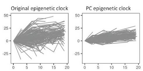
Schematic picture of how the reliability of the epigenetic clock (Y-axis: change in methylation) is improved in the new PC clock in longitudinal data (X-axis: year from baseline) from the research study "the Swedish Adoption Twin Study of Aging" (SATSA).
Credit: Sara Hägg 

Epigenetic clocks based on DNA methylation data are a type of biomarker that is useful for estimating biological age in population-based cohorts. However, usability has been limited by partially unreliable estimates due to this. noise in data. In a new study, we present a solution by introducing a method that reduces the noise in the epigenetic clocks for improved precision in longitudinal analyzes and clinical studies.

Biomarkers for aging can be obtained from cellular, molecular, functional and physiological measurements and used to study biological aging in humans. Many studies use the so-called "epigenetic clocks" based on DNA methylation data to analyze the relationship between biological aging and morbidity and mortality. Although these watches are currently considered to be the best predictors of biological age, they still include technical noise, leading to great variation in the measurements.

In order to make the clocks more useful for longitudinal analyzes and clinical studies, improved precision in the measurements is important. Here we present a method where principal component analysis (PCA) is used to separate noise from age-related signals. In this way, only biologically relevant signals are prioritized and the reliability of the PC watches is much higher compared to the original watches.

Tuesday, August 16, 2022

UF research shows a step toward restoring sea urchins: ‘The lawnmowers of reefs’

Sea urchin
Credit: Josh Patterson 

Coral reef ecosystems are severely threatened by pollution, disease, overharvesting and other factors. For thousands of years, long-spined sea urchins helped keep reefs intact. They eat seaweed, which can kill or seriously damage coral. Without coral, reefs suffer severe consequences, including diminished ability to support fish.

In the mid-1980s, more than 90% of the urchins that crawled the coral reefs in the western Atlantic and Caribbean died for reasons scientists have yet to determine. The population of the long-spined sea urchin – known scientifically as Diadema antillarum -- has been slow to recover on its own. That’s why scientists, including Josh Patterson, are stepping up their efforts to enhance urchin populations.

“You could call these urchins the lawn mowers of the reefs,” said Patterson, a UF/IFAS associate professor of fisheries and aquatic sciences. “They eat fleshy seaweeds that grow out of control on coral reefs and ultimately smother the corals.”

The UF/IFAS restoration ecologist is trying to return more of the urchin to an area that roughly includes the seas off the Florida Keys, Bermuda, the Yucatan Peninsula, Aruba and the Virgin Islands. He’s taken a small step toward the overarching goal of revitalizing the population of the vital echinoderm.

New Technology Sharpens Images of Black Holes

The emission from M87 has now been resolved into a bright, thin ring (orange colormap), arising from the infinite sequence of additional images of the emission region, and the more diffuse primary image, produced by the photons that come directly toward Earth (in blue contours). When viewed at the imaging resolution of the Event Horizon Telescope, the two components blur together. However, by separately searching for the thin ring, it is possible to sharpen the view of M87, isolating the fingerprint of strong gravity.
Credit: Broderick et al. 2022, ApJ, 935, 61

When scientists unveiled humanity’s historic first image of a black hole in 2019 — depicting a dark core encircled by a fiery aura of material falling toward it — they believed even richer imagery and insights were waiting to be teased out of the data.

Simulations predict that, obscured by that bright orange glow, there should exist a thin, bright ring of light created by photons flung around the back of the black hole by its intense gravity.

Now, a team of researchers has combined theoretical predictions and sophisticated imaging algorithms to “remaster” the original imagery of the supermassive black hole at the center of the galaxy M87*, first captured by the Event Horizon Telescope (EHT) in 2019. Their findings, published today in The Astrophysical Journal, are consistent with theoretical predictions and offer new ways to explore these mysterious objects, which are believed to reside at the hearts of most galaxies.

"The approach we took involved leveraging our theoretical understanding of how these black holes look to build a customized model for the EHT data," says Dominic Pesce, a study co-author based at the Center for Astrophysics | Harvard & Smithsonian and member of the EHT collaboration. "Our model decomposes the reconstructed image into the two pieces that we care most about, so that we can study both pieces individually rather than blended together."

Featured Article

Discovery of unexpected collagen structure could ‘reshape biomedical research’

Jeffrey Hartgerink is a professor of chemistry and bioengineering at Rice. Photo Credit: Courtesy of Jeffrey Hartgerink / Rice University Co...

Top Viewed Articles