. Scientific Frontline: Virology
Showing posts with label Virology. Show all posts

Friday, January 6, 2023

Monkeypox viruses remain sensitive to the available drugs

Monkeypox Virus
Illustration Credit: Samuel F. Johanns

The three antiviral drugs commonly used to treat mpox viruses (monkeypox viruses) are also effective against the viruses from the current outbreak. This has been shown in cell culture experiments by scientists at Goethe University Frankfurt/University Hospital Frankfurt and the University of Kent in Canterbury, Great Britain.

The mpox virus is closely related to the smallpox virus (variola virus), which caused large, deadly outbreaks before it was eradicated by vaccination at the end of the 1970s. While the smallpox virus led to very severe disease progression with a death rate of about 30 percent, mpox is milder. Nevertheless, the mortality rate is still about three percent. Particularly at risk of a severe course of the disease are people with a weakened immune system, elderly persons, pregnant women, newborn babies and young children. Until recently, mpox outbreaks only occurred in certain parts of Africa when humans became infected through contact with wild animals, typically rodents such as the Gambian pouched rat and the rope squirrel.

However, in May 2022 a first large mpox outbreak outside Africa was detected; the virus spread solely through human-to-human transmission. This ongoing outbreak has so far reached more than 100 countries and been classified by the World Health Organization (WHO) as a "Public Health Emergency of International Concern".

Thursday, January 5, 2023

Effects of highly pathogenic avian influenza on canids investigated

When the H5N1 HPAI virus (orange) affected a flock of crows in a public garden, it caused a mass die-off of crows. An Ezo red fox and a Japanese raccoon dog were also infected by the H5N1 HPAI virus, the former likely by consuming corpses of the crows, and the latter due to close contact with crow corpses
Illustration Credit: Takahiro Hiono

Researchers at Hokkaido University have revealed the effects of high pathogenicity avian influenza virus infection on an Ezo red fox and a Japanese raccoon dog, linking their infection to a recorded die-off of crows.

High Pathogenicity Avian Influenza (HPAI), commonly known as a type of bird flu, is caused by a group of influenza viruses that affect birds. Humans are very rarely infected by this virus. The most well-known HPAI viral subtype is H5N1, first reported in 1996 for its infection in geese, and then found in humans since 1997. A great amount of time and resources are devoted to monitoring and tracking the spread of HPAI across the globe, due to its disruptive potential on poultry farming—outbreaks are contained by culling exposed and infected flocks.

Thursday, December 15, 2022

Quenchbody Immunosensors Pave the Way to Quick and Sensitive COVID-19 Diagnostics


A new immunosensor based on Quenchbody technology shows great potential as a fast, inexpensive, and convenient tool to detect SARS-CoV-2. Developed by scientists at Tokyo Institute of Technology (Tokyo Tech) and Tokyo Medical and Dental University (TMDU), this highly efficient diagnostic approach will be useful not only for point-of-care testing, but also for high-throughput epidemiological studies of COVID-19 and other emerging infectious diseases.

The double-tagged Quenchbody immunosensor becomes fluorescent when its target antigen—the nucleocapsid protein from SARS-CoV-2—binds at the antigen-binding region of the antibody fragments. This approach is fast, cost-effective, and convenient to use in practice, making it ideal for point-of-care testing as well as batch processing of patient samples. 

The incredibly fast spread of COVID-19 throughout the world brought to light a very important fact: we need better methods to diagnose infectious diseases quickly and efficiently. During the early months of the pandemic, polymerase chain reaction (PCR) tests were one of the most widely used techniques to detect COVID-19. However, these viral RNA-based techniques require expensive equipment and reaction times longer than an hour, which renders them less than ideal for point-of-care testing.

Monday, December 12, 2022

Studies find Omicron related hospitalizations lower in severity than Delta and Pfizer-BioNTech COVID vaccine remains effective in preventing hospitalizations

Photo Credit: Fernando Zhiminaicela

Adult hospitalizations from Omicron-related SARS-CoV-2 (COVID-19) were less severe than Delta and the Pfizer-BioNTech vaccine (also known as Comirnaty and BNT162b2*) remains effective in preventing not only hospitalization, but severe patient outcomes associated with COVID-19, two new research studies have found.

The University of Bristol-led research, funded and conducted in collaboration with Pfizer Inc., as part of AvonCAP, is published in The Lancet Regional Health – Europe.

AvonCAP records adults who are admitted to Bristol’s two hospital Trusts – North Bristol NHS Trust (NBT) and University Hospitals Bristol and Weston NHS Foundation Trust (UHBW) with possible respiratory infection.

In the first paper ‘Severity of Omicron (B.1.1.529) and Delta (B.1.617.2) SARS-CoV-2 infection among hospitalized adults: a prospective cohort study in Bristol, United Kingdom’ researchers assessed whether Delta SARS-CoV-2 infection resulted in worse patient outcomes than Omicron SARS-CoV-2 infection, in hospitalized patients

The study aimed to provide more detailed data on patient outcomes, such as the need for respiratory support.

Friday, December 9, 2022

How a viral toxin may exacerbate severe COVID-19

In a new study, University of California, Berkeley, researchers find that portions of the SARS-CoV-2 “spike” protein, shown in the foreground, can damage the cell barriers that line the inside of blood vessels, contributing to some of COVID-19’s most dangerous symptoms, including acute respiratory distress syndrome (ARDS).
Image Credit: National Institutes of Health

In a new study, University of California, Berkeley, researchers find that portions of the SARS-CoV-2 “spike” protein, shown in the foreground, can damage the cell barriers that line the inside of blood vessels, contributing to some of COVID-19’s most dangerous symptoms, including acute respiratory distress syndrome (ARDS). (National Institutes of Health photo via Flickr)

A study published today in the journal Nature Communications reveals how a viral toxin produced by the SARS-CoV-2 virus may contribute to severe COVID-19 infections.

The study shows how a portion of the SARS-CoV-2 “spike” protein can damage cell barriers that line the inside of blood vessels within organs of the body, such as the lungs, contributing to what is known as vascular leak. Blocking the activity of this protein may help prevent some of COVID-19’s deadliest symptoms, including pulmonary edema, which contributes to acute respiratory distress syndrome (ARDS).

“In theory, by specifically targeting this pathway, we could block pathogenesis that leads to vascular disorder and acute respiratory distress syndrome without needing to target the virus itself,” said study lead author Scott Biering, a postdoctoral scholar at the University of California, Berkeley. “In light of all the different variants that are emerging and the difficulty in preventing infection from each one individually, it might be beneficial to focus on these triggers of pathogenesis in addition to blocking infection altogether.”

SARS-CoV-2 protein caught severing critical immunity pathway

This image shows the SARS-CoV-2 virus's main protease, Mpro, and two strands of a human protein, called NEMO. One NEMO strand (blue) has been cut by Mpro, and the other NEMO strand (red) is in the process of being cut by Mpro. Without NEMO, an immune system is slower to respond to increasing viral loads or new infections. Seeing how Mpro attacks NEMO at the molecular level could inspire new therapeutic approaches. 
Illustration Credit: Greg Stewart/SLAC National Accelerator Laboratory

Over the past two years, scientists have studied the SARS-CoV-2 virus in great detail, laying the foundation for developing COVID-19 vaccines and antiviral treatments. Now, for the first time, scientists at the Department of Energy’s SLAC National Accelerator Laboratory have seen one of the virus’s most critical interactions, which could help researchers develop more precise treatments.

The team caught the moment when a virus protein, called Mpro, cuts a protective protein, known as NEMO, in an infected person. Without NEMO, an immune system is slower to respond to increasing viral loads or new infections. Seeing how Mpro attacks NEMO at the molecular level could inspire new therapeutic approaches.

To see how Mpro cuts NEMO, researchers funneled powerful X-rays from SLAC’s Stanford Synchrotron Radiation Lightsource (SSRL) onto crystallized samples of the protein complex. The X-rays struck the protein samples, revealing what Mpro looks like when it dismantles NEMO’s primary function of helping our immune system communicate.

New findings on how to avert excessive weight loss from COVID-19

Professor Yihai Cao.
Photo Credit: Dr. Muyi Yang.

Losing too much weight when infected with COVID-19 has been linked to worse outcomes. Now, researchers at Karolinska Institutet have discovered that SARS-CoV-2 infection fuels blood vessel formation in fat tissues, thus revving up the body’s thermogenic metabolism. Blocking this process by using an existing drug curbed weight loss in mice and hamsters that were infected with the virus, according to the study published in the journal Nature Metabolism.

“Our study proposes a completely new concept for treating COVID-19 associated weight loss by targeting the blood vessels in the fat tissues,” says Yihai Cao, professor at the Department of Microbiology, Tumor and Cell Biology, Karolinska Institutet, and the study’s corresponding author.

The researchers examined how different types of fat, including brown fat and visceral and subcutaneous white fat, reacted when exposed to SARS-CoV-2 and how it impacted weight in mice and hamsters. They found that the animals lost significant amounts of weight in four days and that this weight loss was preceded by the activation of brown fat and the browning of both types of white fat. These fat tissues also contained more microvessels and high levels of a signaling protein called vascular endothelial growth factor (VEGF), which promotes the growth of new blood vessels.

Corona vaccination also protects people infected with HIV

They represent the study team: Clara Bessen, Carlos Plaza Sirvent, Adriane Skaletz-Rorowski, Anja Potthoff and Agit Simsek (from left).
Photo Credit: RUB, Marquard

A study shows that booster vaccination is particularly useful.

HIV-infected people who receive antiretroviral therapy form antibodies against Sars-Cov-2 after the Corona vaccination with mRNA vaccines. However, your immune response to vaccination is less strong than that of healthy people. The difference is reduced by a third vaccination. These results were achieved by a study with a total of 91 participants, which was carried out by a research team led by Prof. Dr. Ingo Schmitz, head of the Molecular Immunology Department at the Ruhr University in Bochum. The researchers report in the journal Frontiers in Immunology.

Vaccination protection for acquired immunodeficiency

Studies have shown that Sars-Cov-2 vaccines protect otherwise healthy people well against a severe course of Covid-19. It has so far been unclear whether this will also be the case for people with acquired immune deficiency. The research team led by Ingo Schmitz and Dr. Anja Potthoff from the Walk in Ruhr (WIR) Center for Sexual Health and Medicine at the RUB University Hospital included 71 people in her study who are HIV positive and receive antiretroviral therapy. 20 HIV-negative control persons also participated. After the first, second and third vaccinations with the mRNA vaccine from Biontech / Pfizer, they examined the immune response of the participants.

Thursday, December 8, 2022

Common medicine can stop the transmission of HIV infection from mother to child

The risk of transmission of HIV infection from mother to child has been reduced in resource-poor countries.
Photo Credit: Antony Trivet

Antiviral drugs almost completely reduce the risk of mothers passing on HIV infection to their children, even in a low-income country with a high HIV incidence such as Tanzania, according to a new study in The Lancet HIV by researchers from Karolinska Institutet. The discovery raises hopes of achieving the World Health Organization’s goal of eliminating the spread of infection from mother to child.

Only 159 infants were infected

The women were followed for 18 months after giving birth when most of them had stopped breastfeeding. When the researchers examined the mothers’ children, they discovered that only 159 of the more than 13,000 infants had been infected with HIV by the age of 1.5 years. Taking into account the margin of error, this means a risk of 1.4 per cent.

Wednesday, December 7, 2022

New Virus Discovered in Swiss Ticks

Ticks in Switzerland carry a new pathogen: the so-called Alongshan virus. 
Photo Credit: Erik Karits

The Alongshan virus was discovered in China only five years ago. Now researchers at the University of Zurich have found the novel virus for the first time in Swiss ticks. It appears to be at least as widespread as the tickborne encephalitis virus and causes similar symptoms. The UZH team is working on a diagnostic test to assess the epidemiological situation.

Ticks can transmit many different pathogens such as viruses, bacteria, and parasites. Of particular concern are the tickborne encephalitis virus (TBEV), which can cause inflammation of the brain and of the linings of the brain and spinal cord, and bacteria leading to the infectious Lyme disease (borreliosis). The list of pathogens transmitted by ticks continues to increase, also in Switzerland: researchers from the Institute of Virology at the University of Zurich (UZH) have now detected the Alongshan virus (ALSV) for the first time in ticks in Switzerland.

Thursday, December 1, 2022

Experimental COVID-19 Vaccine Offers Long-Term Protection Against Severe Disease

A study involving rhesus macaques at the California National Primate Research Center shows that COVID-19 vaccines given to infant animals protect against lung disease one year after vaccination.
 Photo Credit: CNPRC

Two-dose vaccines provide protection against lung disease in rhesus macaques one year after they were vaccinated as infants, a new study shows. The work, published in Science Translational Medicine Dec. 1, is a follow-up to a 2021 studying showing that the Moderna mRNA vaccine and a protein-based vaccine candidate containing an adjuvant, a substance that enhances immune responses, elicited durable neutralizing antibody responses to SARS-CoV-2 during infancy in preclinical research.

The co-senior authors of the paper are Kristina De Paris, professor of microbiology and immunology at the University of North Carolina at Chapel Hill; Sallie Permar, professor and chair of the Department of Pediatrics at Weill Cornell Medicine; and Koen K.A. Van Rompay, leader of the Infectious Disease Unit at the California National Primate Research at the University of California, Davis. Co-first authors are Emma C. Milligan at the Children’s Research Institute, UNC School of Medicine; and Katherine Olstad at the CNPRC.

To evaluate SARS-CoV-2 infant vaccination, the researchers immunized two groups of eight infant rhesus macaques at the CNPRC at 2 months of age and again four weeks later. Each animal received one of two vaccine types: a preclinical version of the Moderna mRNA vaccine or a vaccine combining a protein developed by the Vaccine Research Center of the National Institute of Allergy and Infectious Diseases (NIAID), with a potent adjuvant formulation. Consisting of 3M’s molecular adjuvant 3M-052 formulated in a squalene emulsion by the Access to Advanced Health Institute (AAHI), the adjuvant formulation stimulates immune responses by engaging receptors on immune cells.

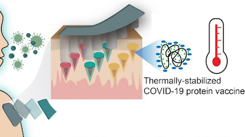
Researchers Develop Strategy to Thermally Stabilize Microneedle Vaccine Technology

Visual of the researcher's microneedle vaccine technology concept.
Illustration Credit: Thahn Nguyen

Researchers use sugar molecules to help eliminate the need for cold-chain storage, a common logistical hurdle for vaccine distribution

Researchers in the Department of Biomedical Engineering —a shared department between the UConn Schools of Dental Medicine, Medicine, and Engineering—unlocked a new strategy using sugar molecules to thermally stabilize their existing microneedle vaccine technology, eliminating the need for cold-chain storage.

Associate Professor Thanh Duc Nguyen from the Departments of Mechanical Engineering and Biomedical Engineering in the School of Engineering, reported this new development in a recent issue of Advanced Materials Technology. The work was led by Dr. Khanh Tran, Nguyen’s former UConn Ph.D. student currently at the Massachusetts Institute of Technology, and Dr. Tyler Gavitt, former UConn Ph.D. student currently at Duke University. Gavitt was a student of Associate Professor Steven Szczepanek in the Department of Pathobiology and Veterinary Science in the College of Agriculture, Health, and Natural Resources at UConn.

Typically, vaccinations against infectious diseases like COVID-19 require multiple painful, expensive and inconvenient injections, including a prime and several booster shots. The UConn researcher’s technology creates a self-administered microneedle patch which could be self-administered and only requires a single-time administration into skin—similar to a nicotine patch—to perform a release profile of vaccines, simulating the effect of multiple injections.

Wednesday, November 30, 2022

Important discovery could help extinguish disease threat to koalas

Retrovirus is more prevalent in New South Wales and Queensland koalas, compared to animals in Victoria and South Australia.
Photo Credit: Jordan Whitt

University of Queensland virologists are a step closer to understanding a mysterious AIDS-like virus that is impacting koala populations differently across state lines.

Dr Michaela Blyton and Associate Professor Keith Chappell from the Australian Institute for Bioengineering and Nanotechnology (AIBN) and School of Chemistry and Molecular Biosciences, have uncovered another piece of the puzzle in their quest to halt the koala retrovirus known as KoRV - a condition strongly associated with diseases that cause infertility and blindness.

“We’ve learned that the retrovirus is far more prevalent in New South Wales and Queensland koalas, compared to the southern populations in Victoria and South Australia,” Dr Blyton said.

“Uncovering crucial patterns like these helps us learn how the disease is evolving, how it’s spreading, and how we can contain the damage through anti-viral medication or koala breeding programs.”

Koala numbers have fallen rapidly over the past decade due to widespread land clearing, climate change induced weather events, and disease.

Dr Blyton’s research has already established the link between KoRV and chlamydia, cystitis and conjunctivitis, which suggests the virus weakens the animal’s immune system.

To track disease-carrying mosquitoes, researchers tag them with DNA barcodes

 The researchers at a field site in Fort Collins, Colorado collecting mosquitoes for analysis.
Photo Credit: Rebekah Kading/Colorado State University

West Nile, Zika, dengue and malaria are all diseases spread by bites from infected mosquitoes. To track the threat of such diseases over large populations, scientists need to know where the mosquitoes are, where they’ve been, and where they might go.

But take it from Rebekah Kading, a Colorado State University researcher who studies mosquito-borne arboviruses: tracking mosquitoes is no easy task. The capture, tagging and release of single mosquitoes – as is commonly done with bats and other disease carriers – would be ridiculous, if not impossible. A common mosquito-tracking technique involves dousing the insects in fluorescent powder and letting them fly away, but the practice is error-prone and unreliable.

Thanks to a collaboration with CSU engineers, Kading and colleagues are introducing a better way to perform mosquito-tracking for disease applications. Their new method, which involves getting larval mosquitoes to eat harmless particles made entirely of DNA and proteins, has the potential to revolutionize how people study mosquito-borne diseases.

The edible mosquito marker particles are the work of Chris Snow, associate professor in the Department of Chemical and Biological Engineering. For the last several years, Snow’s team has been developing microscopic, porous protein crystals that self-assemble from a protein originally found in Camplyobacter jejuni bacteria. Since inventing these very small, non-toxic protein crystals that feature highly precise arrays of pores, Snow’s team has been exploring diverse applications for them, like capturing virus particles to facilitate wastewater testing.

Monday, November 28, 2022

Discovery of antibody structure could lead to treatment for Crimean Congo Hemorrhagic Fever virus

Scott D. Pegan, a professor of biomedical sciences
Photo Source: University of California, Riverside

A research team led by the University of California, Riverside, has discovered important details about how therapeutically relevant human monoclonal antibodies can protect against Crimean Congo Hemorrhagic Fever virus, or CCHFV. Their work, which appears online in the journal Nature Communications, could lead to the development of targeted therapeutics for infected patients.

An emerging zoonotic disease with a propensity to spread, CCHF is considered a priority pathogen by the World Health Organization, or WHO. CCHF outbreaks have a mortality rate of up to 40%. Originally described in Crimea in 1944–1945, and decades later in the Congo, the virus has recently spread to Western Europe through ticks carried by migratory birds. The disease is already endemic in Africa, the Balkans, the Middle East, and some Asian countries. CCHFV is designated as a biosafety level 4 pathogen (the highest level of biocontainment) and is a Category A bioterrorism/biological warfare agent. There is no vaccine to help prevent infection and therapeutics are lacking.

Scott D. Pegan, a professor of biomedical sciences in the UCR School of Medicine, collaborated on this study with the United States Army Medical Research Institute of Infectious Diseases, or USAMRIID, which studies CCHFV because of the threat it poses to military personnel around the world. They examined monoclonal antibodies, or mAbs, which are proteins that bind to antigens — foreign substances that enter the body and cause the immune system to mount a protective response.

In a previous publication, USAMRIID scientists Joseph W. Golden and Aura R. Garrison reported that an antibody called 13G8 protected mice from lethal CCHFV when administered post-infection. They provided Pegan with the sequence information for that antibody, clearing the way for UCR to “humanize” it and conduct further research.

Saturday, November 26, 2022

Blood group can predict risk of contracting viral disease

People with blood type Rh(D) have a higher risk of being infected by parvovirus
Photo Credit: Bartek Kopała

The risk of being infected by parvovirus is elevated in those people who have blood group Rh(D), according to a study published in The Journal of Infectious Diseases by researchers from Karolinska Institutet in collaboration with Octapharma.  

Fifth disease is a viral disease caused by parvovirus. Most often, school-age children are affected with common symptoms such as red blotches on the cheeks that can also spread to the arms and legs. Even adults can become infected, but many do not show any symptoms.  

In a new study, researchers can now demonstrate that the risk of contracting the disease is elevated if the person belongs to the blood group Rhesus D antigen or what is called Rh(D). In addition to the blood typing in the AB0 system, the Rh system is the most common.

Thursday, November 24, 2022

SARS-CoV-2 detection in 30 minutes using gene scissors

Multiplex chip of a Freiburg research team: On this chip, the viral load in the nasal swab and, if necessary, the antibiotic concentration in the blood of COVID-19 patients could be measured simultaneously.
Photo Credit: AG Disposable Microsystems/University of Freiburg

Researchers of the University of Freiburg introduce biosensor for the nucleic acid amplification-free detection of SARS-CoV-2 RNA

CRISPR-Cas is versatile: Besides the controversial genetically modified organisms (GMOs), created through gene editing, various new scientific studies use different orthologues of the effector protein ‘Cas’ to detect nucleic acids such as DNA or RNA.

In its most recent study, the research group headed by microsystems engineer Dr. Can Dincer of the Department of Microsystems2 Engineering, University of Freiburg introduces a microfluidic multiplexed chip for the simultaneous measurement of the viral load in nasal swabs and (if applicable) the blood antibiotic levels of COVID-19 patients.

Rapid test or PCR?

The market launch of rapid antigen test kits has significantly changed the way in which society handles the effects of the pandemic: Individuals suspecting an infection with SARS-CoV-2 can now test themselves at home with kits that are readily available at most drug stores, pharmacies and supermarkets, instead of making an, oftentimes difficult to acquire, appointment for PCR testing, that requires 1 to 3 additional days to receive the result. This convenience is, however, paid for with test sensitivity. This issue became flagrantly apparent during the wave of infections last winter, when the ‘lateral flow devices’ frequently failed to detect infections with the Omicron-variant until after the onset of symptoms. “The trade-off between sensitivity and sample-to-result time could potentially be bridged using our method,” says Midori Johnston, first author of the study, that is now being published in the journal Materials Today.

Wednesday, November 23, 2022

Pocket feature shared by deadly coronaviruses could lead to pan-coronavirus antiviral treatment

Spike glycoprotein structure of SARS-CoV, the coronavirus causing the 2002 outbreak. When linoleic acid is bound, the structure is locked in a non-infectious form. The cryo-EM density, calculated by cloud computing, is shown (left) along with the protein structure (middle). Linoleic acid molecules are colored orange. A zoom-in of the pocket (boxed), conserved in all deadly coronaviruses, is shown
 Illustration Credit: Christiane Schaffitzel and Christine Toelzer, University of Bristol

Scientists have discovered why some coronaviruses are more likely to cause severe disease, which has remained a mystery, until now. Researchers of the University of Bristol-led study, published in Science Advances today [23 November], say their findings could lead to the development of a pan-coronavirus treatment to defeat all coronaviruses—from the 2002 SARS-CoV outbreak to Omicron, the current variant of SARS-CoV-2, as well as dangerous variants that may emerge in future.

In this new study, an international team, led by Bristol's Professor Christiane Schaffitzel, scrutinized the spike glycoproteins decorating all coronaviruses. They reveal that a tailor-made pocket feature in the SARS-CoV-2 spike protein, first discovered in 2020, is present in all deadly coronaviruses, including MERS and Omicron. In striking contrast, the pocket feature is not present in coronaviruses which cause mild infection with cold-like symptoms.

The team say their findings suggest that the pocket, which binds a small molecule, linoleic acid—an essential fatty acid indispensable for many cellular functions including inflammation and maintaining cell membranes in the lungs so that we can breathe properly—could now be exploited to treat all deadly coronaviruses, at the same time rendering them vulnerable to a linoleic acid-based treatment targeting this pocket.

New Omicron subvariant BQ.1.1 resistant to all therapeutic antibodies

The Omicron subvariants BA.1, BA.4, BA.5 as well as Q.1.1 have a high number of mutations in the spike protein. Some of these mutations are escape mutations that allow the virus to escape neutralization by antibodies. In addition, resistance to biotechnologically produced antibodies, which are administered to high-risk patients as a preventive measure or as therapy for a diagnosed SARS-CoV-2 infection, is also developing. Omicron sub-lineage BQ.1.1 is the first variant resistant to all antibody therapies currently approved by the EMA (European Medicines Agency) and/or FDA (US Food and Drug Administration).
Figure Credit: Markus Hoffmann, German Primate Center – Leibniz Institute for Primate Research.

Are the currently approved antibody therapies used to treat individuals at increased risk for severe COVID-19 disease also effective against currently circulating viral variants? A recent study by researchers at the German Primate Center (DPZ) – Leibniz Institute for Primate Research and Friedrich-Alexander University Erlangen-Nürnberg shows that the Omicron sub-lineage BQ.1.1, currently on the rise worldwide, is resistant to all approved antibody therapies. published in the journal Lancet Infectious Diseases.

Tuesday, November 22, 2022

A growing trend of antibody evasion by new omicron subvariants

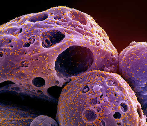
Scanning electron micrograph of a cell (purple) infected with the omicron strain of SARS-CoV-2 virus particles (orange), isolated from a patient sample and colorized.
Image Credit: NIAID/NIH

Three currently circulating omicron subvariants of SARS-CoV-2 – including two that currently make up almost 50% of reported COVID-19 infections in the U.S. – are better at evading vaccine- and infection-generated neutralizing antibodies than earlier versions of omicron, new research suggests.

Scientists tested neutralizing antibodies in blood serum samples from vaccinated and once-boosted or recently infected health care professionals against several subvariants in circulation. Three subvariants stood out for their resistance to the antibody immune response: BQ.1, BQ.1.1 and BA.2.75.2.

BQ.1 and BQ.1.1 are subvariants of the BA.4/5 omicron variants that have been dominating the last few months in the U.S., and each now accounts for about a quarter of current infections, according to the Centers for Disease Control and Prevention (CDC). BA.2.75.2, a mutant of the BA.2 omicron variant, was the best of all variants tested at evading neutralizing antibodies, but currently accounts for only a very small proportion of reported illnesses in the United States.

“In general, the subvariants BQ.1 and BQ.1.1 are much better compared to prior variants at evading the booster-mediated antibody response – the neutralizing antibody titers are clearly much lower. And those two variants are becoming dominant,” said Shan-Lu Liu, senior author the study and a virology professor in the Department of Veterinary Biosciences at The Ohio State University.

Featured Article

Autism and ADHD are linked to disturbed gut flora very early in life

The researchers have found links between the gut flora in babies first year of life and future diagnoses. Photo Credit:  Cheryl Holt Disturb...

Top Viewed Articles