. Scientific Frontline: December 2021

Thursday, December 30, 2021

Elusive atmospheric molecule produced in a lab for the 1st time

Methanediol molecule
Credit: University of Hawaiʻi

The previously elusive methanediol molecule of importance to the organic, atmospheric science and astrochemistry communities has been synthetically produced for the first time by University of Hawaiʻi at Mānoa researchers. Their discovery and methods were published in Proceedings of the National Academy of Sciences on December 30.

Methanediol is also known as formaldehyde monohydrate or methylene glycol. With the chemical formula CH2(OH)2, it is the simplest geminal diol, a molecule which carries two hydroxyl groups (OH) at a single carbon atom. These organic molecules are suggested as key intermediates in the formation of aerosols and reactions in the ozone layer of the atmosphere.

The research team—consisting of Department of Chemistry Professor Ralf Kaiser, postdoctoral researchers Cheng Zhu, N. Fabian Kleimeier and Santosh Singh, and W.M. Keck Laboratory in Astrochemistry Assistant Director Andrew Turner—prepared methanediol via energetic processing of extremely low temperature ices and observed the molecule through a high-tech mass spectrometry tool exploiting tunable vacuum photoionization (the process in which an ion is formed from the interaction of a photon with an atom or molecule) in the W.M. Keck Laboratory in Astrochemistry. Electronic structure calculations by University of Mississippi Associate Professor Ryan Fortenberry confirmed the gas phase stability of this molecule and demonstrated a pathway via reaction of electronically excited oxygen atoms with methanol.

Leveraging Space to Advance Stem Cell Science and Medicine

Arun Sharma, PhD, leads a new research laboratory in the Cedars-Sinai Board of Governors Regenerative Medicine Institute, Smidt Heart Institute and Department of Biomedical Sciences.
Photo by Cedars-Sinai.

The secret to producing large batches of stem cells more efficiently may lie in the near-zero gravity conditions of space. Scientists at Cedars-Sinai have found that microgravity has the potential to contribute to life-saving advances on Earth by facilitating the rapid mass production of stem cells.

A new paper, led by Cedars Sinai and published in the peer-reviewed journal Stem Cell Reports, highlights key opportunities discussed during the 2020 Biomanufacturing in Space Symposium to expand the manufacture of stem cells in space.

Biomanufacturing—a type of stem cell production that uses biological materials such as microbes to produce substances and biomaterials suitable for use in preclinical, clinical, and therapeutic applications—can be more productive in microgravity conditions.

“We are finding that spaceflight and microgravity is a desirable place for biomanufacturing because it confers a number of very special properties to biological tissues and biological processes that can help mass produce cells or other products in a way that you wouldn’t be able to do on Earth,” said stem cell biologist Arun Sharma, PhD, research scientist and head of a new research laboratory in the Cedars-Sinai Board of Governors Regenerative Medicine Institute, Smidt Heart Institute and Department of Biomedical Sciences.

High-resolution lab experiments show how cells ‘eat’

Comert Kural
A new study shows how cell membranes curve to create the “mouths” that allow the cells to consume things that surround them.

“Just like our eating habits basically shape anything in our body, the way cells ‘eat’ matters for the health of the cells,” said Comert Kural, associate professor of physics at The Ohio State University and lead author of the study. “And scientists did not, until now, understand the mechanics of how that happened.”

The study, published recently in the journal Developmental Cell, found that the intercellular machinery of a cell assembles into a highly curved basket-like structure that eventually grows into a closed cage. Scientists had previously believed that structure began as a flat lattice.

Membrane curvature is important, Kural said: It controls the formation of the pockets that carry substances into and out of a cell.

The pockets capture substances around the cell, forming around the extracellular substances, before turning into vesicles – small sacs one-one millionth the size of a red blood cell. Vesicles carry important things for a cell’s health – proteins, for example – into the cell. But they can also be hijacked by pathogens that can infect cells.

But the question of how those pockets formed from membranes that were previously believed to be flat had stymied researchers for nearly 40 years.

“It was a controversy in cellular studies,” Kural said. “And we were able to use super-resolution fluorescence imaging to actually watch these pockets form within live cells, and so we could answer that question of how they are created.

Wednesday, December 29, 2021

Smart sutures to monitor deep surgical wounds

Surgical sutures with an attached electronic module for wireless and battery-free monitoring of deep surgical sites.
Credit: National University of Singapore

Monitoring surgical wounds after an operation is an important step to prevent infection, wound separation and other complications.

However, when the surgical site is deep in the body, monitoring is normally limited to clinical observations or costly radiological investigations that often fail to detect complications before they become life-threatening.

Hard bioelectronic sensors can be implanted in the body for continuous monitoring, but may not integrate well with sensitive wound tissue.

To detect wound complications as soon as they happen, a team of researchers led by Assistant Professor John Ho from the NUS Electrical and Computer Engineering as well as the NUS Institute for Health Innovation & Technology has invented a smart suture that is battery-free and can wirelessly sense and transmit information from deep surgical sites.

These smart sutures incorporate a small electronic sensor that can monitor wound integrity, gastric leakage and tissue micromotions, while providing healing outcomes which are equivalent to medical-grade sutures.

Robots collect underwater litter

The robot of the SeaClear Project is able to detect and collect underwater litter.
Image: The SeaClear Project
Removing litter from oceans and seas is a costly and time-consuming process. As part of a European cooperative project, a team at the Technical University of Munich (TUM) is developing a robotic system that uses machine learning methods to locate and collect waste under water.

Our seas and oceans currently contain somewhere between 26 and 66 million tons of plastic waste, most of which is lying on the seafloor. This represents an enormous threat to marine plants and animals and to the ecological balance of the seas.

But removing waste from the waters is a complex and expensive process. It is often dangerous, too, because the work is generally done by scuba divers. The cleanup operations are also usually limited to the water surface. In the SeaClear Project, a team at TUM is working with eight European partner institutions to develop a robotic system capable of collecting underwater litter.

Tuesday, December 28, 2021

Optimization of mRNA containing nanoparticles

Dr. Aurel Radulescu at the KWS-2 instrument of the Juelich Center for Neutron Science (JCNS) in the research neutron source Heinz Maier-Leibnitz (FRM II) of the Technical University of Munich
Image: Bernhard Ludewig / TUM / FRM II

The research neutron source Hein Maier-Leibnitz (FRM II) at the Technical University of Munich (TUM) is playing an important role in the investigation of mRNA nanoparticles similar to the ones used in the Covid-19 vaccines from vendors BioNTech and Pfizer. Researchers at the Heinz Maier-Leibnitz Zentrum (MLZ) used the high neutron flux available in Garching to characterize various formulations for the mRNA vaccine and thus to lay the groundwork for improving the vaccine's efficacy.

The idea of using messenger RNA (mRNA) as an active ingredient is a brilliant one: The molecule contains the specific blueprint for proteins which are then synthesize by the cell. This makes it generally possible to provide a very wide spectrum of different therapeutically effective proteins.

In the case of the Covid-19 vaccine, these are the proteins of the characteristic spikes on the surface of the Corona virus which are used for vaccination. The proteins are presented on the surface of immune cells; then the human immune system triggers defenses against these foreign proteins and thus against the Corona virus. The mRNA itself is completely broken down after only a few hours, a fact which is advantageous to the safety of these vaccines.

The mRNA has to be packaged appropriately in order to keep it from being broken down on the way to the cell by the ubiquitous enzymes of the human body. This is done using nanoparticles which can consist of a mixture of lipids or polymers.

Engineers bring a soft touch to commercial robotics

Credit: National University of Singapore
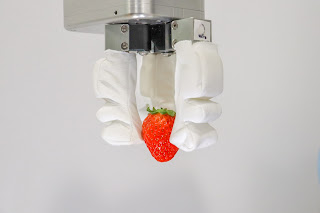
Inspired by the natural dexterity of the human hand, a team of engineers from the National University of Singapore (NUS) has created a reconfigurable hybrid robotics system that is able to grip a variety of objects: from the small, soft and delicate to the large, heavy and bulky. This technology is expected to impact a range of industries, involving food assembly, vertical farming and fast-moving consumer goods packaging, which will progressively automate more of their operations in the coming years.

Led by Associate Professor Raye Yeow from the NUS Department of Biomedical Engineering and the NUS Advanced Robotics Centre, the hybrid robotic grippers use soft, flexible 3D-printed fingers with a reconfigurable gripper base. The robotic innovation is now in the process of being brought to commercial partners under the team’s start-up RoPlus (RO+), comprising NUS researchers Low Jin Huat, Khin Phone May, Chen Chao-Yu and undergraduate student Han Qian Qian.

“An object’s shape, texture, weight and size affect how we choose to grip them. This is one of the main reasons why many industries still heavily rely on human labour to package and handle delicate items,” Assoc Prof Yeow said. “Our hybrid robotic gripper technology revolutionizes traditional pick-and-place tasks by offering advanced capabilities that allow robots to safely interact with delicate items of various shapes, sizes and stiffness, just like the human hand.”

Monday, December 27, 2021

Window to access legal abortion may close before many women know they are pregnant

Credit: Heidi-Ann Fourkiller / SFLORG
More than one in five women experience irregular menstrual cycles that could keep them from learning they are pregnant until it may be too late to access an abortion under some state laws in effect or under consideration, a new study shows.

Researchers from the University of Wisconsin–Madison and National Institutes of Health analyzed anonymized data on 1.6 million menstrual cycles provided by more than 267,000 adults to a cycle-tracking app. According to results published today in the journal Proceedings of the National Academy of Sciences, 22 percent of the people in the study had irregular menstrual cycles that differed in length from one cycle to the next by seven or more days.

“For almost everyone, the first symptom of a pregnancy is a missed period,” says UW–Madison sociology Professor Jenna Nobles, coauthor of the study. “But many people — a large share of the population — have long or highly irregular cycles and could not reasonably learn about their pregnancy in time to seek a legal abortion under laws that set limits at detectable fetal cardiac activity or six weeks.”

Irregular periods are more likely among those with some relatively common medical conditions like Type 2 diabetes, polycystic ovary syndrome and thyroid and other hormone disorders. Hispanic women had greater risk than non-Hispanic white women of experiencing irregular cycles. The age group most like to have cycles of irregular length is 18- to 24-year-olds — also the ages with the highest abortion rates in the United States.

‘Battle of the sexes’ begins in womb as father and mother’s genes tussle over nutrition

Section of mouse fetus and placenta 
Credit: Ionel Sandovici
As the fetus grows, it needs to communicate its increasing needs for food to the mother. It receives its nourishment via blood vessels in the placenta, a specialized organ that contains cells from both baby and mother.

Between 10% and 15% of babies grow poorly in the womb, often showing reduced growth of blood vessels in the placenta. In humans, these blood vessels expand dramatically between mid and late gestation, reaching a total length of approximately 320 kilometers at term.

In a study published today in Developmental Cell, a team led by scientists at the University of Cambridge used genetically engineered mice to show how the fetus produces a signal to encourage growth of blood vessels within the placenta. This signal also causes modifications to other cells of the placenta to allow for more nutrients from the mother to go through to the fetus.

Dr Ionel Sandovici, the paper’s first author, said: “As it grows in the womb, the fetus needs food from its mum, and healthy blood vessels in the placenta are essential to help it get the correct amount of nutrients it needs.

“We’ve identified one way that the fetus uses to communicate with the placenta to prompt the correct expansion of these blood vessels. When this communication breaks down, the blood vessels don’t develop properly and the baby will struggle to get all the food it needs.”

Scientists build new atlas of ocean’s oxygen-starved waters

Oxygen deficient zone intensity across the eastern Pacific Ocean, where copper colors represent the locations of consistently lowest oxygen concentrations and deep teal indicates regions without sufficiently low dissolved oxygen.
Credit: Jarek Kwiecinski and Andrew Babbin

Life is teeming nearly everywhere in the oceans, except in certain pockets where oxygen naturally plummets and waters become unlivable for most aerobic organisms. These desolate pools are “oxygen-deficient zones,” or ODZs. And though they make up less than 1 percent of the ocean’s total volume, they are a significant source of nitrous oxide, a potent greenhouse gas. Their boundaries can also limit the extent of fisheries and marine ecosystems.

Now MIT scientists have generated the most detailed, three-dimensional “atlas” of the largest ODZs in the world. The new atlas provides high-resolution maps of the two major, oxygen-starved bodies of water in the tropical Pacific. These maps reveal the volume, extent, and varying depths of each ODZ, along with fine-scale features, such as ribbons of oxygenated water that intrude into otherwise depleted zones.

The team used a new method to process over 40 years’ worth of ocean data, comprising nearly 15 million measurements taken by many research cruises and autonomous robots deployed across the tropical Pacific. The researchers compiled then analyzed this vast and fine-grained data to generate maps of oxygen-deficient zones at various depths, similar to the many slices of a three-dimensional scan.

Sunday, December 26, 2021

Helium bath splash

Ions packed in a helium nanodroplet remain protected on impact.
Credit: University Innsbruck

While working with helium nanodroplets, scientists at the Department of Ion Physics and Applied Physics led by Fabio Zappa and Paul Scheier have come across a surprising phenomenon: When the ultracold droplets hit a hard surface, they behave like drops of water. Ions with which they were previously doped thus remain protected on impact and are not neutralized.

At the Department of Ion Physics and Applied Physics, Paul Scheier's research group has been using helium nanodroplets to study ions with methods of mass spectrometry for around 15 years. Using a supersonic nozzle, tiny, superfluid helium nanodroplets can be produced with temperatures of less than one degree Kelvin. They can very effectively be doped with atoms and molecules. In the case of ionized droplets, the particles of interest are attached to the charges, which are then measured in the mass spectrometer. During their experiments, the scientists have now stumbled upon an interesting phenomenon that has fundamentally changed their work. "For us, this was a gamechanger," says Fabio Zappa from the nano-bio-physics team. "Everything at our lab is now done with this newly discovered method." The researchers have now published the results of their studies in Physical Review Letters.

Saturday, December 25, 2021

Schizophrenia and bipolar disorder found in recently evolved region of the ‘dark genome’

They say these new proteins can be used as biological indicators to distinguish between the two conditions, and to identify patients more prone to psychosis or suicide.

Schizophrenia and bipolar disorder are debilitating mental disorders that are hard to diagnose and treat. Despite being amongst the most heritable mental health disorders, very few clues to their cause have been found in the sections of our DNA known as genes.

The scientists think that hotspots in the ‘dark genome’ associated with the disorders may have evolved because they have beneficial functions in human development, but their disruption by environmental factors leads to susceptibility to, or development of, schizophrenia or bipolar disorder.

The results are published in the journal Molecular Psychiatry.

“By scanning through the entire genome we’ve found regions, not classed as genes in the traditional sense, which create proteins that appear to be associated with schizophrenia and bipolar disorder,” said Dr Sudhakaran Prabakaran, who was based in the University of Cambridge’s Department of Genetics when he conducted the research, and is senior author of the report.

He added: “This opens up huge potential for new druggable targets. It’s really exciting because nobody has ever looked beyond the genes for clues to understanding and treating these conditions before.”

The researchers think that these genomic components of schizophrenia and bipolar disorder are specific to humans - the newly discovered regions are not found in the genomes of other vertebrates. It is likely that the regions evolved quickly in humans as our cognitive abilities developed, but they are easily disrupted - resulting in the two conditions.

New materials for quantum technologies

Marc A. Wilde investigates materials with special symmetries, such as manganese-silicon, in the laboratory of the TUM chair for Experimental Physics on the Topology of Correlated Systems.
Image: Astrid Eckert / TUM

While conventional electronics relies on the transport of electrons, components that convey spin information alone may be many times more energy efficient. Physicists at the Technical University of Munich (TUM) and the Max Planck Institute for Solid State Research in Stuttgart have now made an important advance in the development of novel materials for such components. These materials may also be the key to quantum computers that are less susceptible to interference.

Hopes ran high when the first representatives of a new class of materials – topological insulators – were discovered some 15 years ago. Researchers predicted that the unique electronic structure of these materials would give rise to special properties on their surface, such as energy-efficient information transmission, which could facilitate the development of novel electronic components in a wide range of applications.

But to date, these possibilities could not be readily modified and controlled in applications. Despite the greatest of efforts, technological exploitation has been a long time coming. This may be about to change thanks to the discovery made by a team headed by Christian Pfleiderer, professorship for the Topology of Correlated Systems at the Technical University of Munich.

Controlled burning of natural environments could help offset our carbon emissions

The finding points to a new method of manipulating the world’s natural capacity for carbon capture and storage, which can also help to maintain natural ecosystem processes. The results are published in the journal Nature Geoscience.

“Using controlled burns in forests to mitigate future wildfire severity is a relatively well-known process. But we’ve found that in ecosystems including temperate forests, savannahs and grasslands, fire can stabilize or even increase soil carbon,” said Dr Adam Pellegrini in the University of Cambridge’s Department of Plant Sciences, first author of the report.

He added: “Most of the fires in natural ecosystems around the globe are controlled burns, so we should see this as an opportunity. Humans are manipulating a process, so we may as well figure out how to manipulate it to maximize carbon storage in the soil.”

Fire burns plant matter and organic layers within the soil, and in severe wildfires this leads to erosion and leaching of carbon. It can take years or even decades for lost soil carbon to re-accumulate. But the researchers say that fires can also cause other transformations within soils that can offset these immediate carbon losses, and may stabilize ecosystem carbon.

Fire stabilizes carbon within the soil in several ways. It creates charcoal, which is very resistant to decomposition, and forms ‘aggregates’ – physical clumps of soil that can protect carbon-rich organic matter at the center. Fire can also increase the amount of carbon bound tightly to minerals in the soil.

Scientists, students will utilize newly launched James Webb Space Telescope for solar system research

The flight mirrors for the James Webb Space Telescope undergo cryogenic testing at NASA Marshall. Credit: Ball Aerospace

In one of the most exciting developments in astronomy in the 21st century, NASA is launching the James Webb Space Telescope (JWST) today—and Northern Arizona University astronomers, planetary astronomers and their students will use the massive observatory to expand their research and advance our understanding of the solar system.

“Webb is NASA’s newest premier space science observatory—destined to be a household name, like its predecessor, Hubble,” NASA announced. “This is an Apollo moment for NASA science—Webb will fundamentally alter our understanding of the universe. It can observe all of the cosmos, from planets to stars to nebulae to galaxies and beyond—helping scientists uncover secrets of the distant universe as well as exoplanets closer to home. Webb can explore our own solar system’s residents with exquisite new detail and search for faint signals from the first galaxies ever made. From new forming stars to devouring black holes, Webb will reveal all this and more.”

The JWST, which NASA calls “a feat of human ingenuity,” is being launched in a global partnership with the European Space Agency and Canadian Space Agency. The mission has evolved over the past 20 years with contributions from thousands of scientists, engineers and other professionals from more than 14 countries and 29 U.S. states, including professor David Trilling, professor Josh Emery and assistant professor Cristina Thomas of NAU’s Department of Astronomy and Planetary Science.

New grafting technique could combat the disease threatening Cavendish bananas

Credit: Steve Hopson
Grafting is the technique of joining the shoot of one plant with the root of another, so they continue to grow together as one. Until now it was thought impossible to graft grass-like plants in the group known as monocotyledons because they lack a specific tissue type, called the vascular cambium, in their stem.

Researchers at the University of Cambridge have discovered that root and shoot tissues taken from the seeds of monocotyledonous grasses - representing their earliest embryonic stages - fuse efficiently. Their results are published today in the journal Nature.

An estimated 60,000 plants are monocotyledons; many are crops that are cultivated at enormous scale, for example rice, wheat and barley.

The finding has implications for the control of serious soil-borne pathogens including Panama Disease, or ‘Tropical Race 4’, which has been destroying banana plantations for over 30 years. A recent acceleration in the spread of this disease has prompted fears of global banana shortages.

“We’ve achieved something that everyone said was impossible. Grafting embryonic tissue holds real potential across a range of grass-like species. We found that even distantly related species, separated by deep evolutionary time, are graft compatible,” said Professor Julian Hibberd in the University of Cambridge’s Department of Plant Sciences, senior author of the report.

Nasal spray to fight COVID-19 heads to clinical trial

The coronavirus in culture.
Credit: Dr Julian Druce VIDRL, Peter Doherty Institute for Infection and Immunity.

An at-home nasal spray treatment for COVID-19 will be put to the test by Melbourne biomedical researchers, as the University of Melbourne and Monash University receive $4.2 million to establish a six-month clinical trial lead by Northern Hospital in collaboration with Oxford University.

Heparin, a widely used blood-thinning drug to treat or prevent blood clots forms the base of the nasal spray treatment that is simple to administer, stable at room temperature and available globally.

Director of Lung Health Research Centre, University of Melbourne Professor Gary Anderson said the spray will be easy to use, with two puffs in each nostril, three times a day.

“Basic science studies revealed that intranasal heparin may be an effective way to prevent COVID-19 infection and spread. COVID-19 first infects cells in the nose, and to do that the virus must bind to Heparan Sulfate on the surface of nasal cells lining the nose,” Professor Anderson said.

“Heparin - the active ingredient in our spray - has a structure that is very similar to Heparan Sulfate, so it behaves as a ‘decoy’ and can rapidly wrap around the virus’s spike protein like a python, preventing it from infecting you or spreading the virus to others.

Friday, December 24, 2021

Lizards offer new possibilities for artificial lungs

Male Brown anole displaying dewlap
When it comes to studying lungs, humans take up all the air, but it turns out scientists have a lot to learn from lizards.

A new study from Princeton University shows how the brown anole lizard solves one of nature’s most complex problems — breathing — with elegant simplicity. Whereas human lungs develop over months and years into baroque tree-like structures, the anole lung develops in just a few days into crude lobes covered with bulbous protuberances. These gourd-like structures, while far less refined, allow the lizard to exchange oxygen for waste gases just as human lungs do. And because they grow quickly by leveraging simple mechanical processes, anole lungs provide new inspiration for engineers designing advanced biotechnologies.

“Our group is really interested in understanding lung development for engineering purposes,” said Celeste Nelson, the Wilke Family Professor in Bioengineering and the principal investigator of a study published in the journal Science Advances. “If we understand how lungs build themselves, then perhaps we can take advantage of the mechanisms mother nature uses to regenerate or engineer tissues.”

While avian and mammalian lungs develop great complexity through endless branching and complicated biochemical signaling, the brown anole lung forms its relatively modest complexity through a mechanical process the authors likened to a mesh stress ball — the common toy found in desk drawers and DIY videos. This study is the first to look at the development of a reptile lung, according to the researchers.

The anole lung starts a few days into development as a hollow, elongated membrane surrounded by a uniform layer of smooth muscle. During development, the lung cells secrete fluid, and as they do so the inner membrane slowly inflates and thins like a balloon. The pressure pushes against the smooth muscle, causing it to tighten and spread apart into fiber bundles that ultimately form a honeycomb-shaped mesh. Fluid pressure continues pushing the stretchy membrane outward, bulging through the gaps in the sinewy mesh and forming fluid-filled bulbs that cover the lung. Those bulges create lots of surface area where the gas exchange occurs.

Research suggests deer could be a possible source of human infection

Douglas Watts, Ph.D., right, professor of biological sciences at The University of Texas at El Paso, and Pedro Palermo, manager of the UTEP Border Biomedical Research Center’s Biosafety Level 3 Infectious Disease Research Program laboratory, are authors of a study that proves for the first time that COVID-19 is present in white-tailed deer in Texas, a finding published recently in Vector-Borne and Zoonotic Diseases.
Photo: J.R. Hernandez / UTEP Marketing and Communications

Research conducted by Douglas Watts, Ph.D., professor of biological sciences at The University of Texas at El Paso, has found COVID-19 present in white-tailed deer in Texas. A report on the discovery was published recently in Vector-Borne and Zoonotic Diseases, a peer-reviewed journal focusing on diseases transmitted to humans by animals.

The UTEP team found the first reported evidence of SARS-CoV-2 infection in deer from Texas, which widens the previously reported geographical range of COVID-19 among deer in the United States, further confirming that infection was common among the species.

“The one thing we know best about SARS-CoV-2 is its unpredictability,” Watts said. “Therefore, the transmission of the virus from infected deer back to humans, while not likely, may be possible.”

While the mechanisms of COVID-19 transmission between humans and animals is still being investigated, the UTEP team’s study suggests that deer should not be neglected as a possible source of SARS-CoV-2 infection among humans as well as domestic and wildlife animals. Watts said subsequent investigations should work to mitigate any risks associated with deer as a possible source of human infection.

COVID-19 infection detected in deer in 9 Ohio locations

Credit: Heidi-Ann Fourkiller SFLORG
Scientists have detected infection by at least three variants of the virus that causes COVID-19 in free-ranging white-tailed deer in six northeast Ohio locations, the research team has reported.

Previous research led by the U.S. Department of Agriculture had shown evidence of antibodies in wild deer. This study, published in Nature, details the first report of active COVID-19 infection in white-tailed deer supported by the growth of viral isolates in the lab, indicating researchers had recovered viable samples of the SARS-CoV-2 virus and not only its genetic traces.

Based on genomic sequencing of the samples collected between January and March 2021, researchers determined that variants infecting wild deer matched strains of the SARS-CoV-2 virus that had been prevalent in Ohio COVID-19 patients at the time. Sample collection occurred before the Delta variant was widespread, and that variant was not detected in these deer. The team is testing more samples to check for new variants as well as older variants, whose continued presence would suggest the virus can set up shop and survive in this species.

The fact that wild deer can become infected “leads toward the idea that we might actually have established a new maintenance host outside humans,” said Andrew Bowman, associate professor of veterinary preventive medicine at The Ohio State University and senior author of the paper.

“Based on evidence from other studies, we knew they were being exposed in the wild and that in the lab we could infect them and the virus could transmit from deer to deer. Here, we’re saying that in the wild, they are infected,” Bowman said. “And if they can maintain it, we have a new potential source of SARS-CoV-2 coming in to humans. That would mean that beyond tracking what’s in people, we’ll need to know what’s in the deer, too.

Experts help to save rare endangered plant from brink of extinction

Ka palupalu o Kanaloa (Photo credit: Zachary Wingert)
Ka palupalu o Kanaloa (Kanaloa kahoolawensis) is one of 10 most critically endangered plants and animals in the world to be impacted by climate change, according to a December 2021 report by the Endangered Species Coalition. The species was declared extinct in the wild in 2015. Over the past several years, experts at the University of Hawaiʻi at Mānoa have brought the Ka palupalu o Kanaloa back from the edge of disappearing from existence.

Ka palupalu o Kanaloa is a densely branched shrub with thin oval leaves and produces large clusters of small white flowers. The plant was historically found to grow on the rocky cliffs of Kahoʻolawe, but fossilized pollen from the species has been found on Oʻahu, Maui, and Kauaʻi. Two Ka palupalu o Kanaloa plants were discovered in 1992 growing on a sea stack off the coast of Kahoʻolawe. Fortunately, seeds were collected from the last two plants before they died in 2015.

Conservation efforts through propagation to save Ka palupalu o Kanaloa were led by Doug Okamoto, a greenhouse technician with UH Mānoa’s Lyon Arboretum, and Anna Palomino from UH Mānoa’s Center for Conservation Research and Training, who is also a Department of Land and Natural Resources Department of Forestry and Wildlife (DLNR-DOFAW) and Plant Extinction Prevention Program horticulturist.

Sugar-coated COVID-19 test takes advantage of coronavirus’ sweet tooth

Ronit Freeman, a UNC-Chapel Hill associate professor of biomedical engineering and applied physical sciences, studies the way coronaviruses enter cells as well as how to simplify the COVID-19 testing process.

Even those tracking each new discovery about the coronavirus and its variants may not be aware of the virus’ sugar cravings.

Researchers at the University of North Carolina at Chapel Hill and University of California San Diego take advantage of the virus’ sweet tooth in the design of a sugar-coated COVID-19 test strip that’s been effective at detecting all known variants of the coronavirus, including delta.

In the next few weeks, researchers will determine if the self-test known as GlycoGrip can detect infections caused by the omicron variant too, said Carolina researcher Ronit Freeman.

“We have turned the tables on the virus by using the same sugar coat it binds to infect cells – to capture it into our sensor,” said Freeman, who published the findings in ACS Central Science.

The test is inspired by the natural biology of epithelial cells – those that are targeted and infiltrated by SARS-CoV-2, the virus that causes COVID-19. These cells are coated with a dense matrix of sugars called the glycocalyx, and it’s this sugar net that the virus exploits to cause infection.

Templating approach stabilizes ‘ideal’ material for alternative solar cells

Artist's impression of formamidinium (FA)-based crystal 
Credit: Tiarnan Doherty
The researchers, from the University of Cambridge, used an organic molecule as a ‘template’ to guide perovskite films into the desired phase as they form. Their results are reported in the journal Science.

Perovskite materials offer a cheaper alternative to silicon for producing optoelectronic devices such as solar cells and LEDs.

There are many different perovskites, resulting from different combinations of elements, but one of the most promising to emerge in recent years is the formamidinium (FA)-based FAPbI3 crystal.

The compound is thermally stable and its inherent ‘bandgap’ – the property most closely linked to the energy output of the device – is not far off ideal for photovoltaic applications.

For these reasons, it has been the focus of efforts to develop commercially available perovskite solar cells. However, the compound can exist in two slightly different phases, with one phase leading to excellent photovoltaic performance, and the other resulting in very little energy output.

“A big problem with FAPbI3 is that the phase that you want is only stable at temperatures above 150 degrees Celsius,” said Tiarnan Doherty from Cambridge’s Cavendish Laboratory, the paper's first author. “At room temperature, it transitions into another phase, which is really bad for photovoltaics.”

Recent solutions to keep the material in its desired phase at lower temperatures have involved adding different positive and negative ions into the compound.

Wednesday, December 22, 2021

ESO telescopes help uncover largest group of rogue planets

This artist’s impression shows an example of a rogue planet with the Rho Ophiuchi cloud complex visible in the background. Rogue planets have masses comparable to those of the planets in our Solar System but do not orbit a star, instead roaming freely on their own. 
Credit: ESO/M. Kornmesser

Rogue planets are elusive cosmic objects that have masses comparable to those of the planets in our Solar System but do not orbit a star, instead roaming freely on their own. Not many were known until now, but a team of astronomers, using data from several European Southern Observatory (ESO) telescopes and other facilities, have just discovered at least 70 new rogue planets in our galaxy. This is the largest group of rogue planets ever discovered, an important step towards understanding the origins and features of these mysterious galactic nomads.

Tuesday, December 21, 2021

Study shows common flower species holds promise for beneficial psychedelic drugs

Thanks to a symbiotic fungus, many species of morning glories contain elements of powerful psychedelic drugs, according to a new Tulane University study published in the journal Communications Biology.

The seeds of the common tropical vine, whose namesake trumpet-like blooms only open in the morning, contain compounds that could be useful for treating mental and physical diseases as well as promoting well-being, said plant and fungal biologist Keith Clay, chairman of the Tulane Department of Ecology and Evolutionary Biology.

Researchers from Tulane, Indiana University and the West Virginia University obtained samples of morning glory seeds from worldwide herbarium collections and screened them for ergot alkaloids, a compound associated with the hallucinogenic drug LSD, but which have also been used for treating migraine headaches and Parkinson’s disease.

Many morning glory species contain high concentrations of bioactive ergot alkaloids that are produced by specialized fungal symbionts passed down from mother plant to offspring through their seeds. Researchers found that one-quarter of over 200 species tested contained ergot alkaloids and were therefore symbiotic.

First model to predict lifetime risk of heart failure

Imagine visiting the doctor, answering a few basic questions and getting an on-the-spot estimate of whether you’ll experience heart failure in the next 30 years.

Such a model now exists, thanks to a new Northwestern Medicine study, which derived and validated the first set of risk prediction models for lifetime risk of heart failure.

The ability to identify who is at greatest risk for heart failure — especially among high-risk young adult populations — will allow physicians to start prevention measures sooner.

“Once someone develops symptoms of heart failure, the window for prevention has closed, which is a missed opportunity, given that the risk of dying in the five years after diagnosis is 50%, similar to a cancer diagnosis,” said corresponding study author Dr. Sadiya Khan, assistant professor of cardiology and epidemiology at Northwestern University Feinberg School of Medicine and a Northwestern Medicine physician.

Prior to this work, no models existed for estimating long-term risk for heart failure. Short-term models exist that estimate heart-failure risk in the next five to 10 years, But those aren’t as effective for young adults who may not develop heart failure until they are older.

Now, for the first time, the model will allow doctors to estimate a person’s risk of developing heart failure in the next 30 years based on their current risk factor levels, such as body mass index, blood pressure, cholesterol, diabetes and smoking status. The scientists are currently working on an online tool that could be used by physicians.

Exquisitely preserved embryo found inside fossilized dinosaur egg

Dinosaur egg and embryo reconstruction
Credit: Julius Csotonyi

A 72- to 66-million-year-old embryo found inside a fossilized dinosaur egg sheds new light on the link between the behavior of modern birds and dinosaurs, according to a new study.

The embryo, dubbed ‘Baby Yingliang’, was discovered in the Late Cretaceous rocks of Ganzhou, southern China and belongs to a toothless theropod dinosaur, or oviraptorosaur. Among the most complete dinosaur embryos ever found, the fossil suggests that these dinosaurs developed bird-like postures close to hatching.

Scientists found the posture of ‘Baby Yingliang’ unique among known dinosaur embryos — its head lies below the body, with the feet on either side and the back curled along the blunt end of the egg. Previously unrecognized in dinosaurs, this posture is similar to that of modern bird embryos.

In modern birds, such postures are related to ‘tucking’ — a behavior controlled by the central nervous system and critical for hatching success. After studying egg and embryo, researchers believe that such pre-hatching behavior, previously considered unique to birds, may have originated among non-avian theropods.

Spray-on coating could make solar panels snow-resistant


In an advance that could dramatically improve the productivity of solar panels in cold climates, a University of Michigan-led team has demonstrated an inexpensive, clear coating that reduced snow and ice accumulation on solar panels, enabling them to generate up to 85% more energy in early testing.

The coating is made chiefly of PVC or PDMS plastic and silicon or vegetable-based oils. It can be sprayed or brushed on in cold weather and, in its current iteration, can keep shedding snow and ice for up to a year.

“Renewable energy is really taking off right now, but snow is a huge problem in northern climates,” said Anish Tuteja, U-M professor of materials science and engineering, who led the study in collaboration with Sandia National Laboratories and the University of Alaska.

“Solar panels might lose 80 or 90% of their generating capacity in the winter. So figuring out a way for them to continue generating energy throughout the year was an exciting challenge,” he said.

While Tuteja’s lab has developed a number of effective ice-shedding coatings in the past, he explains that designing a coating that can passively shed both snow and ice represents a special challenge.

“Ice is relatively dense and heavy, and our previous coatings used its own weight against it,” Tuteja said. “But snow can be 10 times less dense than ice, so we weren’t at all certain that the tricks we use on ice would translate to snow.”

Extinct reptile discovery reveals earliest origins of human teeth, study finds

Infographic showing differentiated teeth
Credit: Dr Suresh Singh
A new extinct reptile species has shed light on how our earliest ancestors became top predators by modifying their teeth in response to environmental instability around 300 million years ago.

In findings published in Royal Society Open Science, researchers at the University of Bristol have discovered that this evolutionary adaptation laid the foundations for the incisor, canine and molar teeth that all mammals - including humans - possess today.

Shashajaia is one of the most primitive members of a group called the Sphenacodontoidea, which includes the famous sail-backed Dimetrodon, and mammal-like reptiles known as therapsids, which eventually evolved into mammals. It is remarkable for its age and anatomy, possessing a very unique set of teeth that set it apart from other synapsids – meaning the animal lineage that mammals belong to - of the time.

Dr Suresh Singh of the School of Earth Sciences explained: “The teeth show clear differentiation in shape between the front and back of the jaw, organized into distinct regions. This is the basic precursor of what mammals have today – incisors and canines up front, with molars in the back. This is the oldest record of such teeth in our evolutionary tree”.

China-wide study will boost understanding of fatal surgical complication

Older patients in hospitals across China took part in a major multi-center open-label randomized clinical trial that showed there was no difference in post-operative delirium in older people with a hip fracture, if they had a general anesthesia, or a regional anesthesia.

Post-operative delirium is a common, serious, neurological, complication where people have a sudden change in their brain functions after an operation. It is more common in older people, and leads to increased death, dementia, post-traumatic stress disorder, a longer length of hospital stay, extra nursing requirements and increased healthcare costs.

People with a hip fracture require an operation to fix the fracture which requires anesthesia - classified as general anesthesia, or regional anesthesia. A general anesthetic technique involves inducing sleep or loss of consciousness through inhaled or intravenous anesthetics. Regional anesthesia involves injecting local anesthetic inside the spine or around the nerves to prevent pain in the leg with the hip fracture.

It was commonly thought that one of the causes of delirium is a general anesthesia. Led by the University of Birmingham and Wenzhou Medical University Second Affiliated Hospital, this randomized trial was the first of its kind in China. 950 older adult took part, from hospitals in Wenzhou, Wuhan, Lishui, Ningbo, Nanchang, and Taizhou.

The trial was set up to provide clinical evidence with a robust methodology, to help medical professionals select appropriate anesthesia for older patients to try and reduce the development of delirium.

Crows keep special tools extra safe

Credit James St Clair

Just like humans, New Caledonian crows are particularly careful when handling their most valuable tools, according to a new study by researchers from the University of St Andrews and the Max Planck Institute of Animal Behavior.

The research, published in the leading scientific journal eLife today (21 December 2021), reveals that crows are more likely to store relatively complex and efficient foraging tools for future use than more basic tools.

New Caledonian crows are renowned for using different types of tools for extracting prey from tree holes and other hiding places. While they firmly hold their tools in the bill during foraging, they need to put them down to eat. This is when crows are at risk of losing their tools by accidentally dropping them or having them stolen by other crows.

In an earlier study, the researchers from the School of Biology at the University of St Andrews had shown that crows keep their tools safe when not needed, using one of two ‘safekeeping’ strategies – they either securely hold them trapped underfoot, or they temporarily insert them into a nearby hole or behind bark. But are crows more careful when handling particularly valuable tools.

New class of galac­tic nebulae disco­vered

Image: Discovery image of the nebula. For this image, 120 individual exposures had to be combined to obtain a total exposure time of 20 hours. The images were taken over several months from Brazil. Credit: Maicon Germiniani

An international team of astronomers led by Stefan Kimeswenger from the Department of Astro and Particle Physics, together with scientific amateurs, has identified a new class of galactic nebulae. This provides an important building block in the understanding of stellar evolution and shows the importance of international collaboration between university research and community science.

For the first time, scientists, starting from a discovery by scientific amateurs, have succeeded in providing evidence for a fully developed shell of a common-envelope-system (CE) – the phase of the common envelope of a binary star system. “Toward the end of their lives, normal stars inflate into red giant stars. Since a very large fraction of stars are in binary stars, this affects the evolution at the end of their lives. In close binary systems, the inflating outer part of a star merges as a common envelope around both stars. However, inside this gas envelope the cores of the two stars are practically undisturbed and follow their evolution like independent single stars,” explains astrophysicist Stefan Kimeswenger. The researchers have now published their results in the journal Astronomy & Astrophysics.

Ground-breaking sensors aboard NASA’s historic space telescope


After NASA launches the James Webb Space Telescope (JWST) on a historic mission this December, scientists anticipate their first glimpse of the most distant objects ever seen in the universe. Technology developed and tested at the University of Hawaiʻi Institute for Astronomy (IfA) and on Maunakea are behind JWST’s ability to gaze deeper into space than ever before.

Sixteen near-infrared (NIR) sensors known as HAWAII-2RGs are part of JWST’s science instruments, enabling it to capture near-infrared light from deep space, far surpassing the capability of NASA’s Hubble Space Telescope. These sensors are the culmination of years of research and development by IfA scientists and engineers. Early prototypes were developed and tested by UH astronomers Don Hall, Klaus Hodapp, and Doug Simons, along with IfA instrumentation engineer Shane Jacobson.

Monday, December 20, 2021

A superstar enzyme is ready for its close-up

This illustration features a cryo-EM “map” of the photosystem II complex. It is a 3D reconstruction, based on two-dimensional cryo-EM images, with different protein subunits of the complex colored individually. Source/Credit: Yale University

A Yale-led team of chemists has unveiled the blueprints for a key enzyme that may contain design principles for a new generation of synthetic solar fuel catalysts.

The research, led by Yale’s Gary Brudvig and Christopher Gisriel, uses cryo-electron microscopy on a microorganism called Synechocystis to get an extreme close-up picture of Photosystem II, the enzyme in photosynthesis that uses water as a solar fuel, enabling researchers to observe how the enzyme works.

The study, which appears in the journal Proceedings of the National Academy of Sciences, was co-authored by researchers from the University of California-Riverside, Boston College, and City University of New York.

Photosynthesis is the mechanism by which plants and certain microorganisms, like Synechocystis, use sunlight to synthesize food from carbon dioxide and water — and fill the atmosphere with oxygen as a byproduct. At the heart of photosynthesis is Photosystem II, an enzyme that oxides water molecules, taking away their electrons to use as fuel.

AI Innovation Incubator to advance artificial intelligence for applied science

Lawrence Livermore National Laboratory (LLNL) has established the AI Innovation Incubator (AI3), a collaborative hub aimed at uniting experts in artificial intelligence (AI) from LLNL, industry and academia to advance AI for large-scale scientific and commercial applications.

LLNL has entered into a new memoranda of understanding with Google, IBM and NVIDIA, with plans to use the incubator to facilitate discussions and form future collaborations around hardware, software, tools and utilities to accelerate AI for applied science. In addition, several existing projects will fall under the AI3 umbrella, including continued work with Hewlett Packard Enterprise (HPE) and Advanced Micro Devices Inc. (AMD) to demonstrate the power of AI and high performance computing (HPC) on the future exascale system El Capitan. This project focuses on innovative, AI-driven cognitive simulation and design optimization methods at unprecedented scales to devise novel approaches to inertial confinement fusion (ICF) experiments at the National Ignition Facility.

Other ongoing projects with AI accelerator/computing companies SambaNova Systems and Cerebras Systems and precision motion company Aerotech, Inc. will be further developed through AI3. More companies, universities and leaders in the AI space are encouraged to consider joining AI3, where early research areas are expected to include advanced material design, 3D printing, predictive biology, energy systems, “self-driving” lasers and fusion energy research.

PPPL unravels a puzzle to speed the development of fusion energy

Yichen Fu, center, lead author of the path-setting paper with co-authors Laura Xing Zhang and Hong Qin.
Photos of Fu and Qin by Elle Starkman/Office of Communications; collage by Kiran Sudarsanan.

Researchers at the U.S. Department of Energy’s (DOE) Princeton Plasma Physics Laboratory have developed an effective computational method to simulate the crazy-quilt movement of free electrons during experimental efforts to harness on Earth the fusion power that drives the sun and stars. The method cracks a complex equation that can enable improved control of the random and fast-moving moving electrons in the fuel for fusion energy.

Fusion produces enormous energy by combining light elements in the form of plasma — the hot, charged gas composed of free electrons and atomic nuclei, or ions, that makes up 99 percent of the visible universe. Scientists around the world are seeking to reproduce the fusion process to provide a safe, clean and abundant power to generate electricity.

Solving the equation

A key hurdle for researchers developing fusion on doughnut-shaped devices called tokamaks, which confine the plasma in magnetic fields, has been solving the equation that describes the motion of free-wheeling electrons as they collide and bounce around. Standard methods for simulating this motion, technically called pitch-angle scattering, have proven unsuccessful due to the complexity of the equation.

Measuring a quantum computer’s power just got faster and more accurate

Sandia National Laboratories has designed a faster, more accurate style of test for quantum computers, such as the one pictured here.
Photo by Bret Latter

What does a quantum computer have in common with a top draft pick in sports? Both have attracted lots of attention from talent scouts. Quantum computers, experimental machines that can perform some tasks faster than supercomputers, are constantly evaluated, much like young athletes, for their potential to someday become game-changing technology.

Now, scientist-scouts have their first tool to rank a prospective technology’s ability to run realistic tasks, revealing its true potential and limitations.

A new kind of benchmark test, designed at Sandia National Laboratories, predicts how likely it is that a quantum processor will run a specific program without errors.

The so-called mirror-circuit method, published today in Nature Physics, is faster and more accurate than conventional tests, helping scientists develop the technologies that are most likely to lead to the world’s first practical quantum computer, which could greatly accelerate research for medicine, chemistry, physics, agriculture and national security.

New Technique Visualizes Every Pigment Cell of Zebrafish in 3D

3D image of melanin in a zebrafish sample captured by micro-computed tomography.
Credit: Spencer R. Katz and Daniel J. Vanselow/Penn State College of Medicine

Researchers have developed a new technique that images every pigment cell of a whole zebrafish in 3D. The work, recently reported in the journal eLife, could help scientists understand the role of melanin in skin cancer.

Melanin is a natural pigment that gives color to the skin, hair, and eyes in humans and animals. Melanin also has implications in melanin-containing cancers, or melanomas, which are typically staged by the depth of penetration in skin.

But studying melanin directly with a conventional microscope is challenging because the pigment blocks light. So Keith C. Cheng, a distinguished professor of pathology, pharmacology and biochemistry, and molecular biology at Penn State College of Medicine, turned to X-ray imaging, which can pass through optically opaque matter like melanin.

To perform the imaging, Cheng partnered with Dula Parkinson, a staff scientist at Berkeley Lab’s Advanced Light Source (ALS), to image two sets of zebrafish samples – one with the normal pigmentation associated with the zebrafish’s characteristic black stripes, and another from a mutant zebrafish line with lighter stripes called golden. Over 15 years ago, Cheng and his lab discovered a key gene implicated in human skin color by studying golden zebrafish. That discovery highlighted the zebrafish’s utility as an animal model of human pigmentation in skin disorders such as albinism or melanoma skin cancer.

Omicron may be significantly better at evading vaccine-induced immunity, but less likely to cause severe disease

As the SARS-CoV-2 virus replicates and spreads, errors in its genetic code can lead to changes in the virus. On 26 Novembe
r 2021, the World Health Organization designated the variant B.1.1.529, first identified in South Africa, a variant of concern, named Omicron. The variant carries a large number of mutations, leading to concern that it will leave vaccines less effective at protecting against infection and illness.

Working in secure conditions, a team led by Professor Ravi Gupta at the Cambridge Institute of Therapeutic Immunology and Infectious Disease, University of Cambridge, created synthetic viruses – known as ‘pseudoviruses’ – that carried key mutations found in the Delta and Omicron strains. They used these to study the virus’s behavior.

The team, which included collaborators from Japan, including Dr Kei Sato of Tokyo University, has released its data ahead of peer review because of the urgent need to share information relating to the pandemic, and particularly the new Omicron variant.

Professor Gupta and colleagues tested the pseudoviruses against blood samples donated to the NIHR COVID-19 BioResource. The blood samples were from vaccinated individuals who had received two doses of either the AstraZeneca (ChAdOx-1) or Pfizer (BNT162b2) vaccines.

On average, Omicron required around a ten-fold increase in the concentration of serum antibody in order to neutralize the virus, compared to Delta. Of particular concern, antibodies from the majority of individuals who had received two doses of the AstraZeneca vaccine were unable to neutralize the virus. The data were confirmed in live virus experiments.

Researchers create artificial cell cortex, a system to study how cells divide

Animal cells are bound by a structure called a cell cortex—and this structure, researchers say, is a bit like a tent.

A tent is constructed of a shell with a zippered opening that controls what can go into and out of the tent. This shell is held up by a system of poles. Similarly, an animal cell cortex is composed of a cell membrane that controls what enters the cell.

The cortex also contains proteins, which help the cell keep its shape. One of these key proteins, called actin, is a polymer with a linear structure—like a tent pole. But unlike a tent, a cell’s cortical proteins aren’t stationary. They move along the cell membrane, freely assembling and moving apart over time, in a process called “cortical excitability.”

When these proteins begin to form wave patterns, it’s a sign that the cell is preparing to divide. But studying this process within the cell membrane is difficult. Now, University of Michigan researchers have developed an approach to study these wave patterns outside of a cell by developing a cell-free artificial cortex.

As a cell prepares to divide, its cell cortex proteins begin to move. First, its cortical proteins form an excitable wave, like spectators performing “the wave” at a football stadium. Second, cortical proteins organize into coherent oscillations, which behave like blinking holiday lights, associating and dissociating with the membrane at regular intervals.
Image credit: Jennifer Landino, A. Miller lab

Vaccine study flips traditional view of product scarcity driving demand

 The first doses of the Pfizer COVID-19 vaccine are administered
to Iowa State University health care employees on
Friday, December 18, 2020, at the Thielen Student Health Center.
Credit: Christopher Gannon/Iowa State University
Anyone who has taken an economics class probably remembers learning about scarcity. The concept of demand outpacing supply applies to the toilet paper shortage at the onset of the COVID-19 pandemic and helps explain how a spike in home-improvement projects last year contributed to skyrocketing lumber prices.

“Previous research on product scarcity shows people will desire something more when it isn’t as easily accessible. Since scarcity signals value, people are willing to make more of an effort or pay more to acquire it,” said Beatriz Pereira, assistant professor of marketing at Iowa State University.

Last year, as COVID-19 cases surged across the U.S., Pereira and a team of researchers knew the initial supply of vaccines would be limited. It seemed like the perfect opportunity to test whether vaccine scarcity drives demand. But the researchers’ newly published findings in Psychology & Marketing reveal the opposite: Participants were less interested in rolling up their sleeves when they thought vaccines were scarce. The researchers point to compassion for the vulnerable as a driving factor.

At the time of the first survey, COVID-19 vaccines were not yet available to the general public.

Over 300 college students were asked to imagine a scenario where manufacturers were working nonstop to produce enough vaccines for everyone, but due to limited supply, priority was being given to people considered high risk. Half of the participants were told that vaccine doses were limited in their area, while the other half were told there were plenty of doses available. The survey then asked both groups of participants the likelihood that they would book a vaccination appointment if their doctor said they could get a shot the following week.

“Interest in booking an appointment dropped by as much as 15% when the participants perceived vaccines as scarce,” said Pereira.

Gum disease increases risk of other illness such as mental health and heart conditions

A University of Birmingham-led study shows an increased risk of patients developing illnesses including mental ill-health and heart conditions if they have a GP-inputted medical history of periodontal (gum) disease.

Experts carried out a first of its kind study of the GP records of 64,379 patients who had a GP-inputted recorded history of periodontal disease, including gingivitis and periodontitis (the condition that occurs if gum disease is left untreated and can lead to tooth loss). Of these, 60,995 had gingivitis and 3,384 had periodontitis. These patients’ records were compared to those of 251,161 patients who had no record of periodontal disease. Across the cohorts, the average age was 44 years and 43% were male, while 30% were smokers. Body Mass Index (BMI), ethnicity and deprivation levels were also similar across the groups.

The researchers examined the data to establish how many of the patients with and without periodontal disease go on to develop cardiovascular disease (e.g., heart failure, stroke, vascular dementia), cardiometabolic disorders (e.g., high blood pressure, Type 2 diabetes), autoimmune conditions (e.g., arthritis, Type 1 diabetes, psoriasis), and mental ill-health (e.g., depression, anxiety and serious mental illness) over an average follow-up of around three years.

From the research, published today in journal BMJ Open, the team discovered that those patients with a recorded history of periodontal disease at the start of the study were more likely to go on and be diagnosed with one of these additional conditions over an average of three years, compared to those in the cohort without periodontal disease at the beginning of the research. The results of the study showed, in patients with a recorded history of periodontal disease at the start of the study, the increased risk of developing mental ill-health was 37%, while the risk of developing autoimmune disease was increased by 33%, and the risk of developing cardiovascular disease was raised by 18%, while the risk of having a cardiometabolic disorder was increased by 7% (with the increased risk much higher for Type 2 diabetes at 26%).

Featured Article

Autism and ADHD are linked to disturbed gut flora very early in life

The researchers have found links between the gut flora in babies first year of life and future diagnoses. Photo Credit:  Cheryl Holt Disturb...

Top Viewed Articles