. Scientific Frontline: Neuroscience
Showing posts with label Neuroscience. Show all posts

Tuesday, January 24, 2023

Propionic acid protects nerve cells and supports their regeneration

Thomas Grüter and Kalliopi Pitarokoili (right) from the study team in St. Josef Hospital.
Photo Credit: RUB, Marquard

Some autoimmune diseases attack the nerves in the arms and legs. Researchers from Bochum are taking a new approach to counteract this damage.

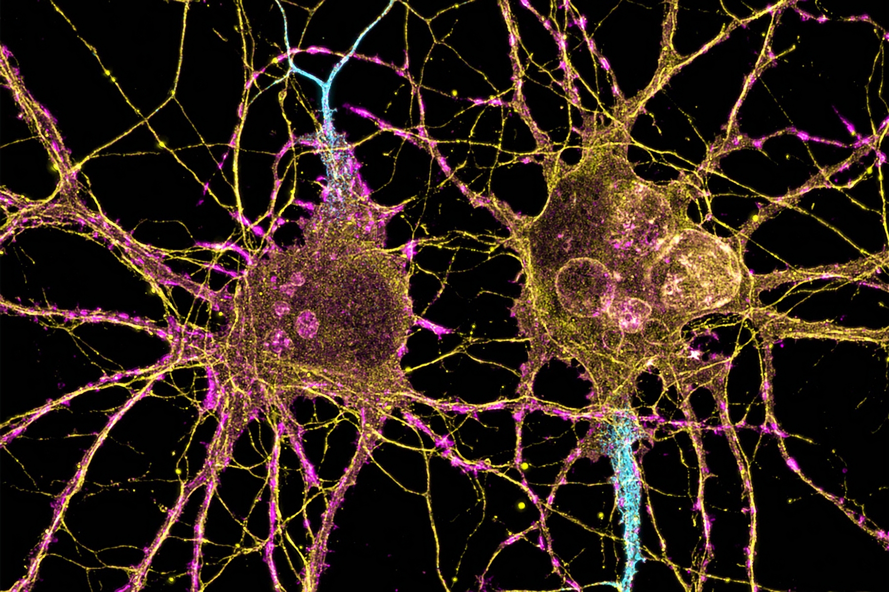
In laboratory tests, researchers from St. Josef Hospital Bochum showed that propionate, the salt of a short-chain fatty acid, can protect nerves and help with their regeneration. The findings could be useful for the treatment of autoimmune diseases that damage nerve cells, such as chronic inflammatory demyelinating polyneuropathy (CIDP). Propionate naturally arises in the intestine when fiber is broken down. In previous studies, a team from the same department from St. Josef Hospital Bochum, clinic of the Ruhr University Bochum, has already proven that people with multiple sclerosis (MS) have a lack of propionate and can benefit from additional propionate intake. Accordingly, the substance could also be useful for patients with CIDP.

A group led by Dr. Thomas Grüter and private lecturer Dr. Kalliopi Pitarokoili from the Neurological University Clinic on St. Josef Hospital (Head of Prof. Dr. Ralf Gold), in the journal Proceedings of the National Academy of Sciences.

Genes Common to Different Species Found to Be Connected to the Development of Depression

Affective disorders, also known as mood disorders, are a group of mental illnesses that involve changes in emotional states.
Photo Credit:: Christopher Lemercier

Russian scientists performed a cross-species analysis of brain gene expression in danio fish, rats and humans to identify new common molecular targets for the therapy of affective disorders of the central nervous system induced by chronic stress. The study was able to identify several key brain proteins that may play important roles in the pathogenesis of affective disorders.

The article was published in the journal Scientific Reports. Affective disorders, also known as mood disorders, are a group of mental illnesses that involve changes in emotional states. They include various forms of depression and mania, psychosis, and increased anxiety. They are widespread because they occur not only as independent mental pathologies, but also as complications of neurological and other somatic diseases.

This fact determines the difficulty of diagnosis: people classify low mood, anxiety and irritability as temporary, situational manifestations. According to statistics, emotional disorders of varying severity occur in 20% of people, but only a quarter of them receive qualified help.

Monday, January 23, 2023

Scientists explain emotional ‘blunting’ caused by common antidepressants

Depression
Photo Credit: Ethan Sykes

According to the NHS, more than 8.3 million patients in England received an antidepressant drug in 2021/22. A widely-used class of antidepressants, particularly for persistent or severe cases, is selective serotonin reuptake inhibitors (SSRIs). These drugs target serotonin, a chemical that carries messages between nerve cells in the brain and has been dubbed the ‘pleasure chemical’.

One of the widely-reported side effects of SSRIs is ‘blunting’, where patients report feeling emotionally dull and no longer finding things as pleasurable as they used to. Between 40-60% of patients taking SSRIs are believed to experience this side effect.

To date, most studies of SSRIs have only examined their short-term use, but, for clinical use in depression these drugs are taken chronically, over a longer period of time. A team led by researchers at the University of Cambridge, in collaboration with the University of Copenhagen, sought to address this by recruiting healthy volunteers and administering escitalopram, an SSRI known to be one of the best-tolerated, over several weeks and assessing the impact the drug had on their performance on a suite of cognitive tests.

In total, 66 volunteers took part in the experiment, 32 of whom were given escitalopram while the other 34 were given a placebo. Volunteers took the drug or placebo for at least 21 days and completed a comprehensive set of self-report questionnaires and were given a series of tests to assess cognitive functions including learning, inhibition, executive function, reinforcement behavior, and decision-making.

Friday, January 20, 2023

How Huntington’s disease affects different neurons

Neuroscientists at MIT have shown that two distinct cell populations in the striatum are affected differently by Huntington’s disease.
Image Credit: Leterrier, NeuroCyto Lab, INP, Marseille, France

In patients with Huntington’s disease, neurons in a part of the brain called the striatum are among the hardest-hit. Degeneration of these neurons contributes to patients’ loss of motor control, which is one of the major hallmarks of the disease.

Neuroscientists at MIT have now shown that two distinct cell populations in the striatum are affected differently by Huntington’s disease. They believe that neurodegeneration of one of these populations leads to motor impairments, while damage to the other population, located in structures called striosomes, may account for the mood disorders that are often see in the early stages of the disease.

“As many as 10 years ahead of the motor diagnosis, Huntington’s patients can experience mood disorders, and one possibility is that the striosomes might be involved in these,” says Ann Graybiel, an MIT Institute Professor, a member of MIT’s McGovern Institute for Brain Research, and one of the senior authors of the study.

Using single-cell RNA sequencing to analyze the genes expressed in mouse models of Huntington’s disease and postmortem brain samples from Huntington’s patients, the researchers found that cells of the striosomes and another structure, the matrix, begin to lose their distinguishing features as the disease progresses. The researchers hope that their mapping of the striatum and how it is affected by Huntington’s could help lead to new treatments that target specific cells within the brain.

Friday, January 6, 2023

The brain’s ability to perceive space expands like the universe

New experiences are absorbed into neural representations over time, symbolized here by a hyperboloid hourglass.
Illustration Credit: Salk Institute

Salk researchers find that neural networks responsible for spatial perception change in a nonlinear manner and may have implications for neurodegenerative disorders like Alzheimer’s disease

Young children sometimes believe that the moon is following them, or that they can reach out and touch it. It appears to be much closer than is proportional to its true distance. As we move about our daily lives, we tend to think that we navigate space in a linear way. But Salk scientists have discovered that time spent exploring an environment causes neural representations to grow in surprising ways.

The findings, published in Nature Neuroscience show that neurons in the hippocampus essential for spatial navigation, memory, and planning represent space in a manner that conforms to a nonlinear hyperbolic geometry—a three-dimensional expanse that grows outward exponentially. (In other words, it’s shaped like the interior of an expanding hourglass.) The researchers also found that the size of that space grows with time spent in a place. And the size is increasing in a logarithmic fashion that matches the maximal possible increase in information being processed by the brain.

Thursday, January 5, 2023

A Theory of Rage

Left: Aditya Nair, Caltech graduate student and study's lead author. Photo Credit: J. Ehlert
Center: Ann Kennedy, Theoretical neuroscientist. Photo Credit: Ann Kennedy
Right: David Anderson, Professor of Biology Photo Credit: Courtesy of David Anderson

Have you ever been cut off while driving and found yourself swearing and laying on the horn? Or come home from a long day at work and lashed out at whoever left the dishes unwashed? From petty anger to the devastating violence we see in the news, acts of aggression can be difficult to comprehend. Research has yielded puzzling paradoxes about how rage works in the brain. But a new study from Caltech, pioneering a machine-learning research technique in the hypothalamus, reveals unexpected answers on the nature of aggression.

The hypothalamus is a brain region linked to many innate survival behaviors like mating, hunting, and the fight-or-flight response. Scientists have long believed that neurons in the hypothalamus are functionally specific—that is, certain groups of neurons correlate to certain specific behaviors. This seems to be the case in mating behavior, where neuron groups in the medial preoptic area (MPOA) of the hypothalamus, when stimulated, cause a male mouse to mount a female mouse. These same neurons are active when mounting behavior occurs naturally. The logical conclusion is that these neurons control mounting in mice.

Wednesday, January 4, 2023

Common Fatty Acid Contributes to Temperature and Pain Sensitivity in Psoriasis Plaques

Photo Credit: Eszter Miller

A common fatty acid found in the Western diet breaks down into compounds that contribute to increased temperature and pain – but not itch – sensitivity in psoriatic lesions. The finding could lead to better understanding of how lipids communicate with sensory neurons, and potentially to improved pain and sensitivity treatments for psoriasis patients.

Linoleic acid is a fatty acid found in vegetable oils, nuts and seeds, and is one of the predominant fatty acids found in the Western diet. Metabolites from linoleic acid – the products formed when the body breaks it down through digestion – play a role in skin barrier function.

“We noticed high levels of two types of lipids derived from linoleic acid in psoriatic lesions,” says Santosh Mishra, associate professor of neuroscience at North Carolina State University and corresponding author of the research. “That led us to wonder whether the lipids might affect how sensory neurons in these lesions communicate. We decided to investigate whether their presence could be related to the temperature or pain hypersensitivity that many psoriasis patients report.”

Sunday, January 1, 2023

Good and bad feelings for brain stem serotonin

An illustration of the facial expression changes in mice following stimulation and inhibition of the median raphe nucleus
Image Credit: Yu Ohmura

New insights into the opposing actions of serotonin-producing nerve fibers in mice could lead to drugs for treating addictions and major depression.

Scientists in Japan have identified a nerve pathway involved in the processing of rewarding and distressing stimuli and situations in mice.

The new pathway, originating in a bundle of brain stem nerve fibers called the median raphe nucleus, acts in opposition to a previously identified reward/aversion pathway that originates in the nearby dorsal raphe nucleus. The findings, published by scientists at Hokkaido University and Kyoto University with their colleagues in the journal Nature Communications, could have implications for developing drug treatments for various mental disorders, including addictions and major depression.

Previous studies had already revealed that activating serotonin-producing nerve fibers from the dorsal raphe nucleus in the brain stem of mice leads to the pleasurable feeling associated with reward. However, selective serotonin reuptake inhibitors (SSRIs), antidepressant drugs that increase serotonin levels in the brain, fail to exert clear feelings of reward and to treat the loss of ability to feel pleasure associated with depression. This suggests that there are other serotonin-producing nerve pathways in the brain associated with the feelings of reward and aversion.

Tuesday, December 20, 2022

Network neuroscience theory best predictor of intelligence

U. of I. Professor Aron Barbey, pictured, and co-author Evan Anderson found that taking into account the features of the whole brain – rather than focusing on individual regions or networks – allows the most accurate predictions of intelligence.     
Photo Credit: Fred Zwicky

Scientists have labored for decades to understand how brain structure and functional connectivity drive intelligence. Researchers report a new analysis offers the clearest picture yet of how various brain regions and neural networks contribute to a person’s problem-solving ability in a variety of contexts, a trait known as general intelligence, researchers report.

They detail their findings in the journal Human Brain Mapping.

The study used “connectome-based predictive modeling” to compare five theories about how the brain gives rise to intelligence, said Aron Barbey, a professor of psychology, bioengineering and neuroscience at the University of Illinois Urbana-Champaign who led the new work with first author Evan Anderson, now a researcher for Ball Aerospace and Technologies Corp. working at the Air Force Research Laboratory.

“To understand the remarkable cognitive abilities that underlie intelligence, neuroscientists look to their biological foundations in the brain,” Barbey said. “Modern theories attempt to explain how our capacity for problem-solving is enabled by the brain’s information-processing architecture.”

Monday, December 19, 2022

Mouse pups cry for help most urgently while active


Mouse pups produce ultrasonic vocalizations, called isolation USVs, when they are separated from the nest. It’s a survival mechanism – baby mice need their parents to regulate their temperature and feed them – that diminishes with age.

But before the USV reflex peters out around 20 days after birth, the rate at which mouse pups cry varies a lot, even within the same individual at the same age, according to Katherine Tschida, the Mary Armstrong Meduski ’80 Assistant Professor of psychology in the College of Arts and Sciences. Exploring this variation, researchers in the Tschida Lab found a link between mouse pup USV rates and their activity levels; the greater amount of body movement, the higher the rate of vocalizations. The connection is important for understanding mouse neural circuitry and development and provides a richer understanding of behavioral differences in mouse models of communication disorders, including autism spectrum disorder (ASD.)

“Rates of Ultrasonic Vocalizations are More Strongly Related Than Acoustic Features to Non-vocal Behaviors in Mouse Pups” was published Dec. 19 in Frontiers in Behavioral Neuroscience. Tschida and doctoral student Nicole Pranic are first authors. Contributions were made by Thomas Cleland, professor of psychology; Chen Yang, programmer and analyst in the Cleland Lab; and by Caroline Kornbrek ’23.

Scientists from NUS and NUHS identify predictive blood biomarker for cognitive impairment and dementia

Prof Barry Halliwell (left) and Dr Irwin Cheah (right), together with their collaborators from the National University Health System, have discovered that low levels of ergothioneine in blood plasma may predict an increased risk of cognitive impairment and dementia.
Photo Credit: National University of Singapore

Identification of elderly persons at risk of developing cognitive impairment and dementia could be made possible by examining ergothioneine levels in the blood

A recent study by a team comprising researchers from the National University of Singapore (NUS) and the National University Health System (NUHS) revealed that low levels of ergothioneine (ET) in blood plasma may predict an increased risk of cognitive impairment and dementia, suggesting possible therapeutic or early screening measures for cognitive impairment and dementia in the elderly.

The research teams were led by Professor Barry Halliwell from the Department of Biochemistry under the NUS Yong Loo Lin School of Medicine and Associate Professor Christopher Chen and Dr Mitchell Lai from the Memory, Ageing and Cognition Centre under NUHS. The results of their most recent study were published in the scientific journal Antioxidants.

Saturday, December 17, 2022

UCLA-developed soft brain probe could be a boon for depression research

 Illustration of the soft probe with aptamer biosensors implanted in the brain.
Illustration Credit: Zhao, et al., 2022

Anyone familiar with antidepressants like Prozac or Wellbutrin knows that these drugs boost levels of neurotransmitters in the brain like serotonin and dopamine, which are known to play an important role in mood and behavior.

It might come as a surprise, then, that scientists still have very little data about the specific relationship between neurotransmitters — chemicals that relay messages from one brain cell to others — and our psychological states. Simply put, monitoring fluctuations of these neurochemicals in living brains has proved a persistent challenge.

Now, for the first time, UCLA scientists have attached nanoscale biochemical sensors, which are tuned to identify specific neurotransmitters, to a soft, implantable brain probe in order to continuously monitor these chemicals in real time. The new brain probe, described in a paper published in ACS Sensors, would allow scientists to track neurotransmitters in laboratory animals — and, ultimately, humans — during their day-to-day activities.

Thursday, December 15, 2022

Frequent genetic cause of late-onset ataxia uncovered by a Quebec-led international collaboration

Photo Credit: whitfieldink

Discovery will improve diagnosis and open treatment possibilities for thousands of people with this debilitating neurodegenerative condition worldwide

A new study published in the New England Journal of Medicine reports the identification of a previously unknown genetic cause of a late-onset cerebellar ataxia, a discovery that will improve diagnosis and open new treatment avenues for this progressive condition.

Late-onset cerebellar ataxias (LOCA) are a heterogeneous group of neurodegenerative diseases that manifest in adulthood with unsteadiness. One to three in 100,000 people worldwide will develop a late-onset ataxia. Until recently, most patients with late-onset ataxia had remained without a genetic diagnosis.

An international team led by Dr. Bernard Brais, a neurologist and researcher at The Neuro (Montreal Neurological Institute-Hospital) of McGill University and Dr. Stephan Züchner of the University of Miami’s Miller School of Medicine, in collaboration with neurologists from the Universities of Montreal and Sherbrooke, studied a group of 66 Quebec patients from different families who had late-onset ataxia for which an underlying genetic cause had not yet been found. Using the most advanced genetic technologies, the team found that 40 (61 per cent) of the patients carried the same novel disease-causing variant in the gene FGF14, making it the most common genetic cause of late-onset ataxia in Quebec. They found that a small stretch of repetitive DNA underwent a large size increase in patients, a phenomenon known as repeat expansion.

Monday, December 12, 2022

Scientists Have Created New Substance to Treat Neurological Disorders

Scientists used a set of 1,2,3-triazole derivatives and modeled the structure of the putative inhibitor.
 Photo Credit: Andrey Fomin

The international team of scientists, including chemists from the Ural Federal University, has developed a substance that may become the basis for drugs that suppress or alleviate a number of neurological disorders. These include, for example, psychosis, schizophrenia, Parkinson's and Huntington's diseases, etc. The scientists reported the development and first results of the study in the Journal of Biomolecular Structure and Dynamics. The study was supported by a grant from the Ministry of Science and Higher Education of the Russian Federation (Project No. 075-15-2020-777).

"We found that the enzyme Phosphodiesterase 10A, which is produced in the body, is directly linked to neurological disorders. If you inhibit this enzyme, you can significantly slow down or even suppress the disease. For this purpose, we used a set of derivatives of 1,2,3-triazole, a pharmacophore whose fragments are contained in many drugs, and modeled the structure of the putative TP-10 inhibitor. We hypothesize that it would have a positive effect on conditions associated with brain dysfunction by reducing the activity of the Phosphodiesterase 10A enzyme. Other inhibitors developed by foreign companies still have no reliable antipsychotic efficacy so far," notes Dhananjay Bhattacherjee, senior researcher at the Department of Organic and Biomolecular Chemistry at UrFU.

Wednesday, December 7, 2022

Using light to manipulate neuron excitability

MIT and Harvard University researchers have devised a way to achieve long-term changes in neuron activity. With their new strategy, they can use light exposure to change the electrical capacitance of the neurons’ membranes, which alters their excitability (how strongly or weakly they respond to electrical signals).
Photo Credit: SFLORG stock image

Nearly 20 years ago, scientists developed ways to stimulate or silence neurons by shining light on them. This technique, known as optogenetics, allows researchers to discover the functions of specific neurons and how they communicate with other neurons to form circuits.

Building on that technique, MIT and Harvard University researchers have now devised a way to achieve longer-term changes in neuron activity. With their new strategy, they can use light exposure to change the electrical capacitance of the neurons’ membranes, which alters their excitability (how strongly or weakly they respond to electrical and physiological signals).

Changes in neuron excitability have been linked to many processes in the brain, including learning and aging, and have also been observed in some brain disorders, including Alzheimer’s disease.

“This new tool is designed to tune neuron excitability up and down in a light-controllable and long-term manner, which will enable scientists to directly establish the causality between the excitability of various neuron types and animal behaviors,” says Xiao Wang, the Thomas D. and Virginia Cabot Assistant Professor of Chemistry at MIT, and a member of the Broad Institute of MIT and Harvard. “Future application of our approach in disease models will tell whether fine-tuning neuron excitability could help reset abnormal brain circuits to normal.”

Monday, December 5, 2022

New findings on neuronal activities in the sensorimotor cortex

Neurons from layer 5 of the motor cortex stained with a fluorescent dye.
Image Credit: Ilka Diester

An interdisciplinary research team at the University of Freiburg has found important clues about the functioning of the sensorimotor cortex. The new findings on neuronal activities in this brain area could be helpful for the further development and use of so-called neuroprostheses. These have an interface with the nervous system and are intended to help compensate for neuronal dysfunctions. "Our results will contribute to the improvement of neuroprosthetic approaches while shortening the training period of patients with prostheses,” says neurobiologist Prof. Dr. Ilka Diester from the Faculty of Biology at the University of Freiburg. The results have just been published in the journal Nature Communications.

Understanding the brain under more natural conditions

The research project also involved the working groups of computer scientist Prof. Dr. Thomas Brox from the University of Freiburg and neuroscientist Prof. Dr. Daniel Durstewitz from the Central Institute of Mental Health in Mannheim. The team found evidence of conserved structures of neuronal activity in the sensorimotor cortex of freely moving rats. The electrophysiological recordings across the entire bilateral sensorimotor cortex allow conclusions about the respective contributions of the premotor, motor and sensory areas. In particular, the researchers found a clear gradient for a contralateral bias, i.e. for movements of the opposite half of the body, from anterior to posterior regions.

Wednesday, November 30, 2022

Better Than a Hole in the Head


Just as blood pressure informs heart health, intracranial pressure (ICP) helps indicate brain health. ICP sensing is the burgeoning focus of Jana Kainerstorfer's biomedical optics lab at Carnegie Mellon University. Her team is working to modernize ICP sensing approaches, which historically have been invasive and risky. Their noninvasive alternatives will ease the risk of infection, pain and medical expenses, as well as present new monitoring capabilities for patients with an array of brain injuries and conditions, from stroke to hydrocephalus.

Investigating pressure levels in the brain is a laborious task for health professionals and hasn't progressed much since the 1960s. Current practice involves drilling a hole into a patient's skull and placing a probe inside for continuous monitoring of ICP levels. It comes with the risk of infection and damaging the brain itself, and while valuable data is to have, ICP measurement is reserved only for the most critical of situations.

"At the core of it, what we've done is build a sensor alternative that doesn't require drilling a hole into the patient's head," said Kainerstorfer, an associate professor of biomedical engineering. "We recently published two papers that explore the use of optical sensors on the forehead for noninvasive ICP monitoring, using near-infrared spectroscopy and diffuse correlation spectroscopy. Both approaches represent huge strides in improving the patient experience and providing better tools to monitor pressure levels in the brain, which can be a key variable in both diagnosis and treatment decisions."

Tuesday, November 29, 2022

Gut Microbes Influence Binge-Eating of Sweet Treats in Mice

Sarkis Mazmanian, Luis B. and Nelly Soux Professor of Microbiology
Photo Credit: Caltech

We have all been there. You just meant to have a single Oreo as a snack, but then you find yourself going back for another, and another, and before you know it, you have finished off the entire package even though you were not all that hungry to begin with.

But before you start feeling too guilty for your gluttony, consider this: It might not be entirely your fault. Now, new research in mice shows that specific gut bacteria may suppress binge eating behavior.

Oreos and other desserts are examples of so-called "palatable foods"—food consumed for hedonistic pleasure, not simply out of hunger or nutritional need. Humans are not alone in enjoying this kind of hedonism: Mice like to eat dessert, too. Even when they have just eaten, they will still consume sugary snacks if available.

The new Caltech study shows that the absence of certain gut bacteria causes mice to binge eat palatable foods: Mice with microbiotas disrupted by oral antibiotics consumed 50 percent more sugar pellets over two hours than mice with gut bacteria. When their microbiotas were restored through fecal transplants, the mice returned to normal feeding behavior. Further, not all bacteria in the gut are able to suppress hedonic feeding, but rather specific species appear to alter the behavior. Bingeing only applies to palatable foods; mice with or without gut microbiota both still eat the same amount of their regular diet. The findings show that the gut microbiota has important influences on behavior and that these effects can be modulated when the microbiota is manipulated.

The brain's immune cells can be triggered to slow down Alzheimer's disease

Joana B. Pereira, researcher at Lund University and Karolinska Institutet who is first author of the study.
Photo Credit: Courtesy of Lund University

The brain's big-eating immune cells can slow down the progression of Alzheimer's disease. This is shown by a study that is now published in Nature Aging.

The brain's own immune cells are called microglia and are found in the central nervous system. They are big eaters that kill viruses, damaged cells and infectious agents they come across. It has long been known that microglial cells can be activated in different ways in several neurological diseases such as Alzheimer's and Parkinson's diseases. Depending on how they are activated, they can both drive and slow disease development. Researchers from Lund University and Karolinska Institutet have now shown that a certain type of activation of the microglial cells triggers inflammatory protective mechanisms in the immune system:

“Most people probably think that inflammation in the brain is something bad and that you should inhibit the inflammatory system in case of illness. But inflammation doesn't just have to be negative”, says Joana B. Pereira, researcher at Lund University and Karolinska Institutet who is first author of the study.

Thursday, November 24, 2022

A brain circuit underpinning locomotor speed control

Zebrafish Photo Credit: Petr Kuznetsov

Researchers at Karolinska have uncovered how brain circuits encode the start, duration and sudden change of speed of locomotion. The study is published in the journal Neuron.

Important findings

By exploiting the relative accessibility of adult zebrafish, combined with a broad range of techniques, the researchers can now reveal two brain circuits that encode the start, duration and sudden change in locomotor speed.

The brain circuits represent the initial step in the sequence of commands coding for the onset, duration, speed and vigor of locomotion. The two command streams revealed here, with their direct access to the spinal circuits, allow the animal to navigate through their environment by grading the speed and strength of their locomotor movements, while at the same time controlling directionality. These mechanisms in adult zebrafish can be extrapolated to mammalian model systems.

Mapping connectivity

The next step will be to map the connectivity between these brain circuits and those in the spinal cord driving locomotion.

Hopefully, the circuit revealed in the study can guide designing novel therapeutic strategies aimed at restoring motor function after traumatic spinal cord injury.

The study was financed by The Swedish Research Council, Knut and Alice Wallenberg Foundation, The Swedish Brain Foundation.

Source/Credit: Karolinska Institutet | Charlotte Brandt

ns112422_01

Featured Article

Autism and ADHD are linked to disturbed gut flora very early in life

The researchers have found links between the gut flora in babies first year of life and future diagnoses. Photo Credit:  Cheryl Holt Disturb...

Top Viewed Articles