. Scientific Frontline: Medical
Showing posts with label Medical. Show all posts

Friday, April 14, 2023

Drug form of traditional Chinese medicine compound improved survival of mice with brain tumors

Indirubin is a natural product present in indigo plants and the active ingredient of the traditional Chinese medicine Dang Gui Long Hui Wan, which is used to treat chronic diseases.
Photo Credit: Courtesy of Brown University

A new study shows how a drug made from a natural compound used in traditional Chinese medicine works against malignant brain tumors in mice, creating a promising avenue of research for glioblastoma treatment.

In the study, published in Cell Reports Medicine, researchers showed how a formulation of the compound, called indirubin, improved the survival of mice with malignant brain tumors. They also tested a new formulation that was easier to administer, taking the potential pharmaceutical approach one step closer to clinical trials with human participants.  

“The interesting thing about this drug is that it targets a number of important hallmarks of the disease,” said Sean Lawler, lead author, associate professor of pathology and laboratory medicine, and researcher at the Legorreta Cancer Center of Brown University. “That's appealing because this type of cancer keeps finding ways around individual mechanisms of attack. So, if we use multiple mechanisms of attack at once, perhaps that will be more successful.”

Curtin researchers map genetic signature of precursor to liver cancer

Photo Credit: Julia Koblitz

Researchers at Curtin University have identified the genetic signature of pre-malignant liver cells, offering potentially significant implications for the almost 3,000 Australians diagnosed with the deadly cancer each year.

The study, published in the prestigious journal Cell Genomics, found that quantifying pre-malignant liver cells in patients with liver disease could help determine their future risk of developing liver cancer.

First author Dr Rodrigo Carlessi, from the Curtin Medical School and the Curtin Health Innovation Research Institute, said the discovery had the potential to save lives by changing how chronic liver disease patients are staged and monitored based on their cancer risk.

“The research used cutting-edge technology to identify the molecular fingerprint of thousands of genes, one cell at a time,” Dr Carlessi said.

“During this process, we discovered the genetic signature and its diagnostic value, which was subsequently confirmed in several hundred individual patient liver samples.

Thursday, April 13, 2023

AI Tool Predicts Colon Cancer Survival, Treatment Response

New AI tool accurately predicts both overall survival and disease-free survival after colorectal cancer diagnosis.
Image Credit: bodymybody

A new artificial intelligence model designed by researchers at Harvard Medical School and National Cheng Kung University in Taiwan could bring much-needed clarity to doctors delivering prognoses and deciding on treatments for patients with colorectal cancer, the second deadliest cancer worldwide.

Solely by looking at images of tumor samples — microscopic depictions of cancer cells — the new tool accurately predicts how aggressive a colorectal tumor is, how likely the patient is to survive with and without disease recurrence, and what the optimal therapy might be for them.

Having a tool that answers such questions could help clinicians and patients navigate this wily disease, which often behaves differently even among people with similar disease profiles who receive the same treatment — and could ultimately spare some of the 1 million lives that colorectal cancer claims every year.

Tuesday, April 11, 2023

Modified Botox gives long-term pain relief after nerve injury without side effects

A single injection of the elongated Botox could relieve pain for months without risk of paralysis or addiction
Photo Credit: Mufid Majnun

A modified form of Botox could give long-term pain relief to patients with chronic nerve injury pain, according to a new study.

A team of scientists from the Universities of Sheffield, Reading and University College London (UCL) and US-based biopharmaceutical company Neuresta have created a new, elongated botulinum neurotoxin which can alleviate chronic pain without risk of paralysis or addiction. 

Chronic pain is extremely difficult to manage, and currently available drugs are limited by dangerous side effects. Opioids like morphine and fentanyl are the gold standard for short-term pain relief but they cannot effectively treat chronic pain due to the risk of addition, abuse and overdose. 

Findings of the new study, published in the journal Life Science Alliance, show that a single injection of the precisely engineered botulinum neurotoxin provides long-lasting relief in mice models, without adverse effects.

The team, led by Professor Bazbek Davletov, Chair of Biomedical Science, and Research Associate Charlotte Leese from the University of Sheffield, developed a new way of rebuilding Botox by using elements of Clostridium botulinum and created a biopharmaceutical with new properties, without unwanted toxic effects. 

Neutrons for better vaccines against multidrug resistant germs

Dr. Jia-Jheng Kang prepares measurements for the vaccines at the KWS-2 sample site.
Photo Credit: Bernhard Ludewig, FRM II / TUM

Neutrons from the Research Neutron Source Heinz Maier-Leibnitz (FRM II) can be used to explore the structure of biomolecules. The most recent success: the precise analysis of a promising vaccine against multidrug resistant germs.

Bacteria which are resistant to all conventional antibiotics cause more than a million deaths each year. Consequently, researchers around the world are searching for new therapeutic approaches to combat these pathogens. Two years ago, an international team in Grenoble identified an active ingredient suitable for the production of a vaccine against multidrug resistant bacteria Pseudomonas aeruginosa. The vaccine has in the meantime been successfully tested on mice.

"As with many new vaccines, in this case the active ingredient is embedded in liposomes. The exact characterization and understanding of these nanoscopic biomolecules is a key factor in the development and optimization of future vaccines," says Dr. Marco Maccarini, biophysicist at the French National Centre for Scientific Research (CNRS). Together with experts at the TIMC laboratory of the Université Grenoble Alpes (UGA) and at the FRM II he has successfully analyzed the structure of the candidate vaccine against Pseudomonas aeruginosa.

Global study finds some women experience heavier menstrual flow after COVID-19 vaccination

Analyses showed a small increase in the percentage of participants who experienced greater total bleeding quantity following the first COVID-19 vaccine dose compared with an unvaccinated comparison group.
Photo Credit: Obi

A new international study finds that women vaccinated for COVID-19 have a slightly higher risk for a heavier period after vaccination.

The study, led by Oregon Health & Science University reproductive health services researcher Blair Darney, Ph.D., M.P.H., and physician-scientist Alison Edelman, M.D., M.P.H., published in the British Journal of Obstetrics and Gynaecology. These findings build on prior work from the same research team that first identified an association between COVID-19 vaccines and menstrual cycle changes.

While there is a growing body of evidence demonstrating that COVID-19 vaccination is associated with a small increase in cycle length, other disturbances such as bleeding quantity are less well known. This study aimed to estimate the effect of COVID-19 vaccination on menstrual bleeding quantity among individuals with normal menstrual cycles.

“Menstruation is a routine bodily function and a key indicator of overall health, so it’s crucial that we understand the scope of this issue among the global population,” said Edelman, one of the study’s lead authors. “The more we can understand about these reported changes, the more effectively we’re able to counsel individuals about what to expect with a COVID-19 vaccine and how to make an informed decision about getting vaccinated.”

Prior treatments influence immunotherapy response in advanced melanoma

Photo Credit: Ivan Samkov

Research led by scientists at UCLA Jonsson Comprehensive Cancer Center found that responses to a type of immunotherapy called PD-1 checkpoint blockade in patients with advanced melanoma depended on whether or not they had previously received another immunotherapy – CTLA-4 blockade – as well as other factors.

Their findings, based on analysis of seven data sets generated over the past decade, which included results of tumor biopsies from more than 500 patients, are published in Cancer Cell.

“In our large set of data, features that have been used to predict response to anti-PD-1 checkpoint blockade therapy – often called biomarkers –related to the presence of certain immune cell types in the tumor and the genetic profile of the tumors themselves were modified by a patient’s treatment history,” said lead author Katie Campbell, PhD, a postdoctoral fellow in hematology/oncology at UCLA Jonsson Comprehensive Cancer Center.

When a patient is diagnosed with advanced melanoma, they usually are treated with immune therapies like anti-PD-1 blockade and anti-CTLA-4 blockade, in combination or alone. By blocking different proteins that diminish the effectiveness of T cells, these checkpoint inhibitors enhance the body’s immune response to cancer.  

Thursday, March 30, 2023

Allies or enemies of cancer: the dual fate of neutrophils

Neutrophils infiltrating tumors are heterogeneous and different neutrophil types can have opposing effects on cancer progression. The image shows artistic rendering of a lung tumor nodule (in blue) infiltrated by various neutrophil types (shown in green, orange and red) including some (in red) that are expanded by immunotherapy and are required for tumor elimination.
Illustration Credit: © Mate Kiss, Evangelia Bolli and Mikael Pittet

An international team including scientists from the UNIGE and Harvard has discovered a new type of immune cell whose action is essential for the success of immunotherapies.

Why do cancer immunotherapies work so extraordinarily well in a minority of patients, but fail in so many others? By analyzing the role of neutrophils, immune cells whose presence usually signals treatment failure, scientists from the University of Geneva (UNIGE), from Harvard Medical School, and from Ludwig Cancer Center have discovered that there is not just one type of neutrophils, but several. Depending on certain markers on their surface, these cells can either promote the growth of tumors, or fight them and ensure the success of a treatment. By boosting the appropriate factors, neutrophils could become great agents of anti-tumor immunity and reinforce the effects of current immunotherapies. These results can be read in Cell.

Lab-made antibodies offer potential cure for yellow fever

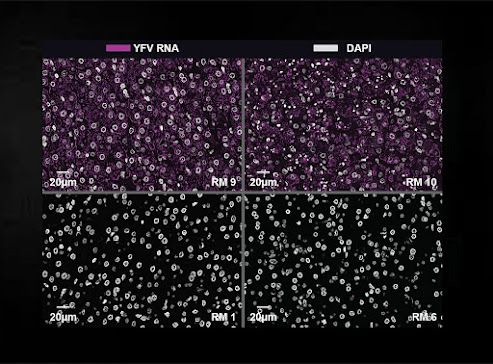
Captured through a microscope, this enlarged image illustrates how yellow fever virus (purple coloring) is below detectable levels in the blood of research animals given a monoclonal antibody after being exposed to the virus (bottom squares). By comparison, yellow fever virus is clearly visible in the blood of research animals that didn’t receive a monoclonal antibody (top squares). This research suggests lab-made antibodies may be able to cure people who get sick with yellow fever, a disease for which there is no approved treatment.
Image Credit: Oregon Health & Science University

New research from Oregon Health & Science University and collaborators indicates lab-made antibodies may be able to cure people infected with yellow fever, a virus for which there is no treatment.

The natural immune response to invading pathogens normally involves making protective proteins called antibodies. A study published in Science Translational Medicine suggests that a single monoclonal antibody infusion can strengthen the body’s fight against yellow fever.

In the study, the yellow fever virus was undetectable in all animals that received monoclonal antibody infusions after being exposed to the virus.

“Two monoclonal antibodies that we evaluated completely removed all signs of infection from research animals,” said the study’s corresponding author, Ben Burwitz, Ph.D., associate professor at OHSU’s Vaccine and Gene Therapy Institute and affiliate associate professor at OHSU’s Oregon National Primate Research Center.

Wednesday, March 29, 2023

Retinoic Acid Could Be Key to Preventing Gut Infections

Brian Sheridan conducts research on CD8 T cells to investigate immune responses with the hope of laying groundwork for new therapies and vaccines. 
Photo Credit: John Griffin, Stony Brook University

A team of scientists from the Renaissance School of Medicine (RSOM) at Stony Brook University have identified a distinct role of retinoic acid, a metabolite of vitamin A, during the immune response of the gut. This finding, detailed in a paper published in the Journal of Experimental Medicine, and highlighted in a broader piece in the journal, could help lead to ways to control the retinoic acid response and therefore could be used as a therapy or for vaccine development against infection or even to treat GI tumors.

Led by Brian Sheridan, associate professor in the Department of Microbiology and Immunology and the Center for Infectious Diseases, the study involves basic research that centers on unraveling the factors that control the generation of cytotoxic memory CD8 T cells, which are an important arm of the body’s anti-pathogen immune response as they kill pathogen-infected cells and produce anti-pathogen cytokines. In fact, memory CD8 T cells provide long-lived and frontline protection at barrier tissues, highlighting their importance in vaccine design.

To date, scientists have known that retinoic acid in the gut-draining lymph nodes promotes effector CD8 T cell migration to the intestines, enhancing the immune response. Additionally, vitamin A deficiency is associated with increased infections and poor vaccine efficiency.

Tuesday, March 28, 2023

Early study shows cones in retinal degeneration, thought to be dormant, may retain visual function

“While the sensitivity of the cones was about 100-1000 fold less than normal, we were surprised to find that that the drop-off in sensitivity for the ganglion cells that project to the brain was much less,” said senior author Alapakkam Sampath.
Photo Credit: Miranda Scalabrino

New UCLA research in mice suggests that “dormant” cone photoreceptors in the degenerating retina are not dormant at all, but continue to function, producing responses to light and driving retinal activity for vision.  

The cells in the retina that produce the visual experience are rods and cones. Rods are active in dim light and cones in daylight. Mutations in rods that cause them to die trigger most inherited retinal degeneration. Cones can remain alive after nearly all the rods die, but they retract key parts of the cells and appear “dormant.”  

But while past literature suggested that dormant cells were not functional, and earlier attempts to record from them revealed no light-driven activity, the new study indicates for the first time that the cells are still viable. Furthermore, downstream signals recorded from the retina show that visual processing is not as compromised as may be expected. The authors say their findings demonstrate that therapeutic interventions to protect these cells, or enhance their sensitivity, have the capability to preserve nearly normal daytime vision. 

Monday, March 27, 2023

Components of Cytoskeleton Strengthen Effect of Sex Hormones

Super-high-resolution microscopic image of a cell that has been exposed to the sex hormone dihydrotestosterone. The androgen receptor (violet) and actin (green) are visible in the cell nucleus. Both molecules are stained with fluorescent dyes and thus made visible. The individual structures made visible (right) are only 200 nanometers (200 millionths of a millimeter) small.
Image Credit: Julian Knerr/University of Freiburg

Researchers from Freiburg and Kiel discover that actin acts in the cell nucleus and is partly responsible for the expression of male sexual characteristics

Steroid hormones, to which belong sex hormones like estrogen or testosterone, are important signaling molecules and are responsible among other things for controlling female and male phenotypic sex differentiation. They act by binding to receptor molecules that switch on and off the activity of hormone-dependent genes. Researchers at the University of Freiburg and Kiel University Hospital have discovered that components of the cytoskeleton are critically involved in this process. The findings are relevant for the diagnosis of medical conditions and the study of diseases and cancers in which steroid hormones play important roles. The study was published in the renowned journal Nature.

The new research findings show that filamentous actin, a component of the cytoskeleton, interacts with the androgen receptor directly in the cell nucleus and strengthens its effect. The androgen receptor mediates the signals of sex hormones for male sex development but also promotes the progression of prostate cancer.

Friday, March 24, 2023

Revolutionary discovery for blood clotting

Treatment with soluble GPV prevents the formation of a vascular-closing thrombus in an experimental mouse model for thrombosis formation (right). On the left is a vascular-closing thrombus of an untreated mouse.
Image Credit: Sarah Beck / University Hospital Würzburg

The platelet glycoprotein V is an important switching point for hemostasis and thrombus formation. This new finding could have great clinical potential.

If our blood vessels are injured by cuts or abrasions or bruises, it is vital that the bleeding is stopped and the wound closed. In technical terms, this process is called hemostasis. This consists of two processes: the hemostasis, in which platelets (platelets) attach to the wound edges, form a plug and temporarily seal the injury. And the blood clotting or coagulation cascade, in which long fibers are formed from fibrin, which together with the platelets seal the wound firmly.

However, if fibrin is formed in excess, for example in chronic wounds, vascular occlusions, so-called thrombosis, can occur. Strict regulation of fibrin formation is therefore important. However, how coagulation is limited has not yet been fully understood.

In an international project coordinated by Würzburg University Medicine, researchers have now deciphered a central regulatory mechanism for fibrin formation and derived new therapeutic approaches from it. The results are released in the renowned journal Nature Cardiovascular Research.

A readily available dietary supplement may reverse organ damage caused by HIV and antiretroviral therapy

Photo Credit: Courtesy of MitoQ

MitoQ, a mitochondrial antioxidant that is available to the public as a diet supplement, was found in a mouse study to reverse the detrimental effects that HIV and antiretroviral therapy (ART) have on mitochondria in the brain, heart, aorta, lungs, kidney and liver.

The researchers used a molecular method to measure the ratio of human and murine mitochondrial (mtDNA) to nuclear DNA (ntDNA) ratio, a measure of mitochondrial dysfunction. Reduction in this ratio reflects mitochondrial dysfunction. Compared to uninfected mice, HIV infected mice treated with ART had mitochondrial dysfunction in the human immune cells in the brain, heart, liver, lungs, and gut. ART itself also affected mitochondrial function in mouse heart cells. When treated with MitoQ for 90 days, HIV infected mice had reduced mitochondrial dysfunction in organs compared to HIV infected mice on ART.

Mitochondria are the key cell structures that are important for the smooth function of organs such as the brain, heart, liver and kidney. HIV causes a chronic state of inflammation and immune dysfunction that contribute to damage to organs. The reasons for this are unclear, but it is known that mitochondrial dysfunction contributes to organ damage and is present in chronic HIV. There are no therapies for HIV associated diseases that affect organs such as the brain, heart and liver.

Thursday, March 23, 2023

Can Artificial Intelligence Predict Spatiotemporal Distribution of Dengue Fever Outbreaks with Remote Sensing Data?

Image Credit: Sophia University
Full Size Image

Researchers train machine learning model with climatic and epidemiology remote sensing data to predict the spatiotemporal distribution of disease outbreaks

Cases of dengue fever and other zoonotic diseases will keep increasing owing to climate change, and prevention via early warning is one of our best options against them. Recently, researchers combined a machine learning model with remote sensing climatic data and information on past dengue fever cases in Chinese Taiwan, with the aim of predicting likely outbreak locations. Their findings highlight the hurdles to this approach and could facilitate more accurate predictive models.

Outbreaks of zoonotic diseases, which are those transmitted from animals to humans, are globally on the rise owing to climate change. In particular, the spread of diseases transmitted by mosquitoes is very sensitive to climate change, and Chinese Taiwan has seen a worrisome increase in the number of cases of dengue fever in recent years.

Like for most known diseases, the popular saying “an ounce of prevention is worth a pound of cure” also rings true for dengue fever. Since there is still no safe and effective vaccine for all on a global scale, dengue fever prevention efforts rely on limiting places where mosquitoes can lay their eggs and giving people an early warning when an outbreak is likely to happen. However, thus far, there are no mathematical models that can accurately predict the location of dengue fever outbreaks ahead of time.

Attack from the intestine

After an operation, bacteria can enter the organism from the intestine. Combat special cells of the immune system that are located in the liver.
Illustration Credit: Mercedes Gomez de Agüero

Darmbacteria are more common triggers of complications after surgery. This is shown by a new study by research teams from Würzburg and Bern. A solution to this problem could come from the liver.

German hospitals carried out almost 16 million operations in 2021. In Switzerland there are around 1.1 million. Even if the actual procedure is going well, it is not uncommon for a wound infection to occur afterwards, which can have dramatic consequences for those affected. In extreme cases, such infections are fatal.

A new study now shows that the causes of these infections are in a large part of the cases bacteria from the patient's intestine itself. To do this, the intestine does not even have to be injured during the operation. In this way, too, these pathogens overcome the intestinal barrier postoperatively and spread throughout the body through the blood and lymphatic pathways. They can be stopped by special immune cells that patrol all organs, including the liver.

Clues to the cause of chronic gut pain

Professor Stuart Brierley
Photo Credit: Courtesy of Flinders University

New insights into chronic gut pain offer hope for improved treatments for irritable bowel syndrome and anxiety treatment.

A research team led by Flinders University Professor Stuart Brierley, based at the SA Health and Medical Research Institute, with Nobel Laureate Professor David Julius, Professor Holly Ingraham and Dr James Bayrer at the University of California San Francisco, has shown evidence of a specific pathway of cells and nerves linking the gut to the brain that may be responsible for the chronic gut pain.

Chronic gut pain is commonly experienced by 11% of the global population currently living with irritable bowel syndrome (IBS) and associated psychological conditions, including anxiety and depression.

Described in a new article in Nature, the team used genetic and pharmacologic tools in pre-clinical models to manipulate signals between gut epithelial cells and associated nerve fibers to determine how this pathway stimulates chronic gut pain and anxiety.

Researchers create artificial enzyme for fast detection of disease-related hormone in sweat

Photo Credit: Courtesy of Oregon State University

Researchers in the Oregon State University College of Engineering have developed a handheld sensor that tests perspiration for cortisol and provides results in eight minutes, a key advance in monitoring a hormone whose levels are a marker for many illnesses including various cancers.

Findings were published in the journal ACS Applied Materials & Interfaces. The material and sensing mechanism in the new device could be easily engineered to detect other specific hormones, the researchers say – for example, progesterone, a key marker for women’s reproductive health and pregnancy outcomes.

“We took inspiration from the natural enzymes used in blood glucose meters sold at pharmacies,” said Larry Cheng, associate professor of electrical engineering and computer science. "In glucose meters, specific enzymes are applied to an electrode, where they can capture and react with glucose molecules to generate an electrical signal for detection. However, finding natural enzymes for cortisol detection is not straightforward, and natural enzymes are prone to instability and have a short lifespan.

Wednesday, March 22, 2023

Pregnant women diagnosed with cancer don’t get the emotional support they need due to research gap

Photo Credit: Lucas Mendes

Support for pregnant women diagnosed with cancer is limited because of insufficient research into the specific emotional consequences and needs associated with a diagnosis at this time, according to a new report from the University of Surrey.

Researchers have also found that pregnant women diagnosed with cancer often delay seeking medical help because they believe their symptoms are due to natural changes in their body.

In the most comprehensive study of its kind, researchers from Surrey, in collaboration with the charity Mummy’s Star, reviewed causes of psychosocial issues (distress, depression, and anxiety) affecting pregnant women diagnosed with cancer and what supportive care is available to them and their partners.

Emory researchers shine light on how stress impacts women's hearts

Stress has significant impact on women's heart health, research has found
Photo Credit: Engin Akyurt

Right in the middle of women’s history month, it’s staggering to think back on how recently women and their hearts began to be taken seriously by the scientific community. As legendary Emory cardiologist Nanette Wenger, MD, wrote in a 2016 American College of Cardiology article: “Although heart disease is the number one killer of women, cardiovascular disease was really thought of as a man’s disease until the last few decades.” 

In the not-so-distant past, Wenger added, “Women who came into the emergency room with chest pains were told they had a stomach problem or that they were imagining the pain and had emotional problems, so they were sent home.” 

Thankfully, following down the path first carved out by pioneers like Wenger, there are researchers and physicians like Viola Vaccarino, MD, PhD, who have continued to build a data-backed case for the fact that women are very much not just making things up. 

Vaccarino, the Wilton Looney Professor of Cardiovascular Research at Rollins’ Department of Epidemiology and faculty member in the Division of Cardiology, is the principal investigator of a prospective study funded by the National Institutes of Health (NIH) looking at sex differences in bodily responses to mental stress and subsequent cardiovascular events among young and middle-aged patients who survived a heart attack at Emory University.  

Featured Article

Autism and ADHD are linked to disturbed gut flora very early in life

The researchers have found links between the gut flora in babies first year of life and future diagnoses. Photo Credit:  Cheryl Holt Disturb...

Top Viewed Articles