. Scientific Frontline: Microbiology
Showing posts with label Microbiology. Show all posts

Tuesday, November 1, 2022

Better understanding of the development of intestinal diseases

Dr. Bahtiyar Yilmaz, First author
Department for BioMedical Research, University of Bern, and Department of Visceral Surgery and Medicine, Inselspital, Bern University Hospital
Credit:  zvg / Courtesy of Bahtiyar Yilmaz

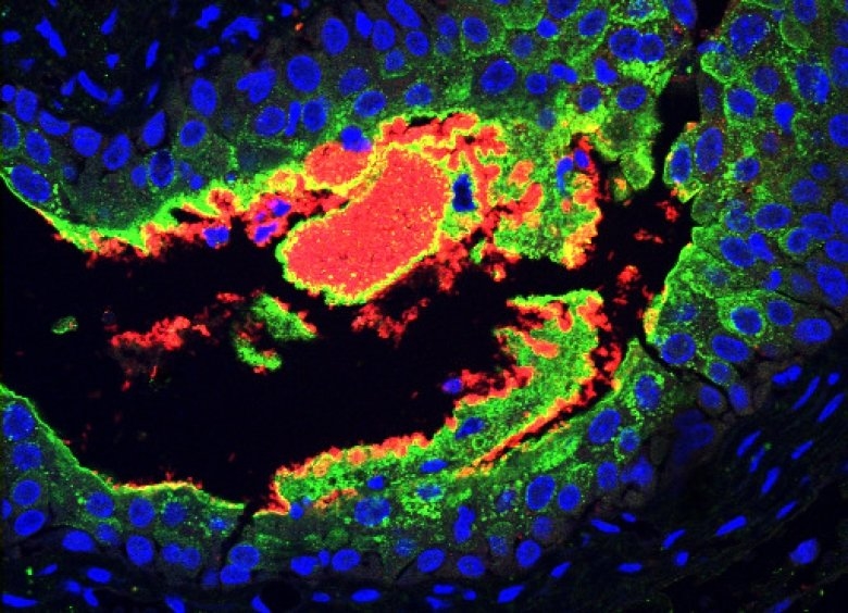
Bacteria in the small intestine adapt dynamically to our nutritional state, with individual species disappearing and reappearing. Researchers at the University of Bern and University Hospital Bern have now been able to comprehensively study the bacteria of the small intestine and their unique adaptability for the first time. The findings contribute to a better understanding of intestinal diseases such as Crohn's disease or celiac disease and to the development of new therapeutic approaches.

Humans have just as many microbes in their microbiota as there are cells in the body, and most of these are in the large intestine (colon). They are an important part of our ‘digestion’ because they can harvest energy from many foods that evade our digestive enzymes. Unfortunately, whilst it is easy to collect fecal samples, it has been largely impossible to study the lower small intestine because this can only be reached during a surgical operation or after purging the intestinal contents to allow safe passage of an endoscope.

The small intestinal microbiome has remained almost “terra incognita” within the human gastrointestinal tract, despite the fact that the small intestine is essential for life and it absorbs 90% of all our calories. Researchers led by Andrew Macpherson and Bahtiyar Yilmaz from the Department for Biomedical Research at the University of Bern and the University Clinic for Visceral Surgery and Medicine at the Inselspital have now been able to examine the intestinal bacteria of the human small intestine in a simple and innovative way to show how they support the digestive process by reacting dynamically to the human nutritional status. While the gut bacteria (microbiota) of the large intestine remain relatively stable throughout life, those in the small intestine have been shown to be very unstable: they largely disappear when we fast overnight and reappear when we eat in the morning. These findings are important for a better understanding of the development of intestinal diseases such as celiac disease or Crohn's disease. The study is published in the journal Cell Host and Microbe.

Monday, October 31, 2022

Bulking Up to Beat Bacteria

The inhibitor-binding site of the wild-type MexB pump. (a) The crystal structure of the inhibitor ABI-PP bound to the MexB trimer. Three MexB monomers are shown in green, blue, and red, representing the access, binding, and extrusion monomer, respectively. ABI-PP is shown as a yellow space-filling model. (b) A close-up view of the inhibitor binding site. The substrate translocation pathway is shown as a solid gray surface. The proximal and distal binding pockets are indicated in green and blue circles, respectively. The inhibitor binding pit is shown as a red surface. The ABI-PP molecule is represented as a yellow stick model. (c) A detailed view of the inhibitor-binding site. Carbon atoms of ABI-PP are indicated in yellow while amino acid residues are indicated in green. The classification of these amino acids is shown on the right side of the panel.
Image Credit: 2022 Yamasaki et al., Spatial Characteristics of the Efflux Pump MexB Determine Inhibitor Binding, Antimicrobial Agents and Chemotherapy

The medical profession is in the midst of losing an arms race. Bacterial antibiotic resistance doesn’t just threaten our ability to treat infection but our ability to carry out any treatment where infection is a risk. This includes a raft of life-saving surgeries ranging from coronary bypass operations to organ transplantation. In fact, the number of new antimicrobials being developed is declining each year. Understanding how bacteria resist the influence of antibiotics is essential to winning this arms race: it is time to make up ground.

In a study published this month in Antimicrobial Agents and Chemotherapy, researchers at Osaka University have produced new insights into the structure of a particular bacterial protein known as an efflux pump. This protein is involved in antibiotic resistance and its structure influences the ability of drugs to target it.

Wednesday, October 26, 2022

Study Identifies Key T Cells for Immunity Against Fungal Pneumonia

 GM-CSF+ and IL-17A+ lineages of T cells are instrumental in controlling many fungal and bacterial infections and implicated in autoimmune pathology. This study shows that GM-CSF expressing Tc17 cells are necessary for mediating fungal vaccine immunity without augmenting pathology.
Credit: Som Nanjappa

Researchers at the University of Illinois College of Veterinary Medicine have demonstrated in a mouse model that a specific type of T cell, one of the body’s potent immune defenses, produces cytokines that are necessary for the body to acquire immunity against fungal pathogens. This finding could be instrumental in developing novel, effective fungal vaccines.

Despite vaccines being hailed as one of the greatest achievements of medicine, responsible for controlling or eradicating numerous life-threatening infectious diseases, no vaccines have been licensed to prevent or control human fungal infections.

This lack proved especially deadly during the COVID-19 pandemic. In countries where steroids were widely used to suppress inflammation of the lungs, COVID-19 patients with preexisting conditions such as uncontrolled diabetes showed a greater likelihood of developing lethal fungal infections.

Saturday, October 22, 2022

Aging, Frailty, and our Microbiomes

Photo Credit: Magda Ehlers

We humans tend to think we live independently, capable of ensuring our own health and wellbeing. As researchers are increasingly aware, however, our microbiomes—the trillions of microbes that live on and within us—play central roles in our health and susceptibility to different diseases. And as we age, our microbiomes change too, with important health implications over time.

Jackson Laboratory (JAX) Associate Professor Julia Oh, Ph.D., studies the microbiome, particularly the microbes that colonize the skin. While prior research has explored the gut microbiome in the context of aging, to date there has been little insight into the changes that occur in other microbial communities of our body, like the mouth and skin. To further investigate, Oh and her team collaborated with UConn Center on Aging Professors Julie Robison, Ph.D., and George Kuchel, M.D., to study the microbiome of the skin, oral, and gut of older adults compared to younger adults.

Because of the unique design of their study, where they sampled frail older adults inhabiting skilled nursing facilities as well as community-dwelling older adults, they found that the greatest microbiome differences between the groups were associated with increased frailty, not chronological age. A second surprising finding was that microbiome differences between cohorts were most pronounced in the skin, rather than the gut or mouth. Moreover, the skin harbored the greatest number of potential risk factors for infectious disease. The researchers presented their findings in “Associations of the skin, oral and gut microbiome with aging, frailty and infection risk reservoirs in older adults,” published in Nature Aging.

“This was an extraordinary multidisciplinary effort between our clinical and research team at UConn Center on Aging and The Jackson Laboratory for Genomic Medicine,” says Oh. “We believe this exciting study is an important step to understanding how the microbiome contributes to aging and chronic diseases, in turn allowing us to identify potential interventional targets to improve health across lifespan.”

Wednesday, October 19, 2022

Methane-Eating ‘Borgs’ Have Been Assimilating Earth’s Microbes

A digital illustration inspired by methane-eating archaea and the Borgs that assimilate them
Credit: Jenny Nuss/Berkeley Lab

In Star Trek, the Borg are a ruthless, hive-minded collective that assimilate other beings with the intent of taking over the galaxy. Here on nonfictional planet Earth, Borgs are DNA packages that could help humans fight climate change.

Last year, a team led by Jill Banfield discovered DNA structures within a methane-consuming microbe called Methanoperedens that appear to supercharge the organism’s metabolic rate. They named the genetic elements “Borgs” because the DNA within them contains genes assimilated from many organisms. In a study published today as the cover item in the journal Nature, the researchers describe the curious collection of genes within Borgs and begin to investigate the role these DNA packages play in environmental processes, such as carbon cycling.

First contact

Methanoperedens are a type of archaea (unicellular organisms that resemble bacteria but represent a distinct branch of life) that break down methane (CH4) in soils, groundwater, and the atmosphere to support cellular metabolism. Methanoperedens and other methane-consuming microbes live in diverse ecosystems around the world but are believed to be less common than microbes that use photosynthesis, oxygen, or fermentation for energy. Yet they play an outsized role in Earth system processes by removing methane – the most potent greenhouse gas – from the atmosphere. Methane traps 30 times more heat than carbon dioxide and is estimated to account for about 30 percent of human-driven global warming. The gas is emitted naturally through geological processes and by methane-generating archaea; however, industrial processes are releasing stored methane back into the atmosphere in worrying quantities.

Tuesday, October 18, 2022

Algae Could be Instrumental in Making Human Exploration of Mars Possible

 A researcher working in UNLV geoscientist Elisabeth "Libby" Hausrath's lab.
Credit: University of Nevada, Las Vegas

While the world is marveling over the first images and data now coming from NASA’s Perseverance rover mission seeking signs of ancient microscopic life on Mars, a team of UNLV scientists is already hard at work on the next step: What if we could one day send humans to the Red Planet?

There’s a lot to consider when sending people, though. Human explorers, unlike their rover counterparts, require oxygen and food, for starters. It also takes about six to nine months — both ways — just in travel time. And then there’s the air itself. Martian air is roughly 98% carbon dioxide (Earth’s is a fraction of 1% for comparison) and the air temperature averages an extremely frigid -81 degrees.

It’s these challenges that UNLV geochemist and NASA Mars 2020 team scientist Libby Hausrath and postdoctoral researcher Leena Cycil, a microbial ecologist, are exploring. And a big part of the answer? Algae.

“Extremophilic algae” are types of algae known for their ability to thrive in extreme environments such as high-altitude snowy mountains or hypersaline lakes. These algae love carbon dioxide and can use it to produce oxygen. They also are edible, dense with nutrients, and grow quickly. Extremophiles’ helpful characteristics allow them to grow in some of the most inhospitable environments on Earth, possibly even in conditions similar to Mars.

Attack on 2 fronts leads ocean bacteria to require carbon boost

The study is the first to observe these complex interactions under the ocean surface: photosynthetic bacteria simultaneously infected with viruses and floating in the presence of organisms, called protists, that eat them. Photo Credit: Matt Hardy

The types of ocean bacteria known to absorb carbon dioxide from the air require more energy – in the form of carbon – and other resources when they’re simultaneously infected by viruses and face attack from nearby predators.

Viruses are abundant in the ocean, and research now suggests that marine viruses have beneficial functions, including helping to drive carbon absorbed from the atmosphere to permanent storage on the ocean floor. When viruses infect other microbes in that environment (and anywhere, in fact), the interaction results in creation of entirely new organisms called “virocells.”

In this new study, researchers worked with cyanovirocells – cyanobacteria that absorb carbon and release oxygen through photosynthesis that have been infected with viruses. The analysis of changes in the infected bacteria’s gene activation and metabolism under lab conditions designed to mimic nature hints at an intriguing possibility: The dual threat of viral infection and drifting among hungry predator microbes might lead cyanovirocells to take in more carbon.

Obesity and biological sex may make individuals more vulnerable to COVID-19

A new West Virginia University study suggests obesity may impair the ability to fight off SARS-CoV-2, the virus that causes COVID-19, in a sex-dependent manner.
Credit: WVU Illustration/Graham Curry

A new animal study from Katherine Lee, a researcher with the West Virginia University School of Medicine, investigates why individuals with obesity may have a particularly difficult time fending off SARS-CoV-2, the virus that causes COVID-19. Specifically, female obese mice experienced worse disease symptoms, showing the importance of both obesity and biological sex in COVID-19 outcomes.

Lee’s findings appear in the journal iScience.

Obesity dramatically increases someone’s risk of being hospitalized, placed on a ventilator or dying due to COVID-19. Considering that about two out of every five Americans are obese, that risk is far from negligible.

“No human is 100% healthy in every respect,” said Lee, a doctoral student in the Department of Microbiology, Immunology and Cell Biology. “There are always going to be little differences in the way our bodies function and those changes can ultimately affect the ways we respond to everything. So, I think as soon as we start incorporating those differences and changes — metabolic diseases and preexisting conditions — into our work, we can learn more about how vaccines and therapeutics might be more or less effective in these people.”

Thursday, September 22, 2022

Gut Microbiomes Help Bears with Very Different Diets Reach the Same Size

Photo credit: National Park Service.

A recent study of the gut microbiome of Alaskan brown bears (Ursus arctos) shows that the microbial life in bears’ guts allows them to achieve comparable size and fat stores while eating widely different diets. The work sheds light on the role of the gut microbiome in supporting health in wild omnivores.

“We think of bears as having simple digestive tracts, so it’s easy to slip into thinking that they therefore have simple gut microbiomes,” says Erin McKenney, co-author of the study and an assistant professor of applied ecology at North Carolina State University. “But this study shows there can be tremendous diversity in the gut microbiomes between individual bears, and that this variation can be very important to the physical condition of these animals.”

“For example, the amount of fat that bears are able to store is absolutely critical to the health of wild populations,” says Grant Hilderbrand, co-author of the study and associate regional director for resources for the National Park Service in Alaska. “If female bears are able to reach levels where 19-20% of their body mass in the autumn is fat, they’ll reproduce. And knowing that they can take different dietary paths to reach those fat levels is a valuable insight.”

For this study, researchers collected fecal samples from 51 adult brown bears in three national parks: Katmai National Park and Preserve, Lake Clark National Park and Preserve, and Gates of the Arctic National Park and Preserve.

Tuesday, September 20, 2022

Mysterious soil virus gene seen for first time

Crystals of the soil virus AMG product (chitosanase) at 400x magnification. Individual crystals were cryo-cooled in liquid nitrogen before being exposed to the powerful SSRL X-rays beams for structure analysis.
Credit: Clyde Smith/SLAC National Accelerator Laboratory

In every handful of soil, there are billions of bacteria, fungi, and viruses, all working to sustain the cycle of life. Understanding how these microorganisms interact with one another helps scientists analyze soil health, soil carbon and nutrient cycling, and even the ways in which dead insects decompose.

Soil viruses contain genes that appear to have some metabolic function, but they are clearly not required for normal viral replication. These genes are called auxiliary metabolic genes (AMGs) and they produce proteins, some of which are enzymes that have a variety of functions. Until now, scientists have wondered whether some AMG proteins play a role in critical soil processes, like carbon cycling. To find out more about soil AMGs, researchers determined the atomic structure of a protein that is expressed by a particular AMG.

Specifically, researchers irradiated fragile crystallized protein samples with high-brightness X-rays generated by the Stanford Synchrotron Radiation Lightsource’s (SSRL) Beam Line 12-2 at the Department of Energy’s (DOE) SLAC National Accelerator Laboratory. The X-rays struck the proteins within the crystal samples, revealing their molecular structures and a bit of the mystery behind their makeup.

AMGs do not, like many viral genes, help a virus replicate. Instead, they encode a variety of proteins, each with their own predicted function. The AMG that was expressed was a putative enzyme that plays a key role in how soils process and cycle carbon in the biosphere.

Scientists Unveil New System for Naming Majority of the World’s Microorganisms

Fluorescent-stained bacteria (pink) and archaea (green) from near-boiling water from Great Boiling Spring in Gerlach, Nevada.
Photo credit: Jeremy Dodsworth.

What’s in a name? For microorganisms, apparently a lot.

Prokaryotes are single-celled microorganisms - bacteria are an example - that are abundant the world over. They exist in the oceans, in soils, in extreme environments like hot springs, and even alongside and inside other organisms including humans.

In short, they’re everywhere, and scientists worldwide are working to both categorize and communicate about them. But here’s the rub: Most don’t have a name.

Less than 0.2% of known prokaryotes have been formally named because current regulations – described in the International Code of Nomenclature of Prokaryotes (ICNP) – require new species to be grown in a lab and freely distributed as pure and viable cultures in collections. Essentially, to name it you have to have multiple physical specimens to prove it.

In an article published in the journal Nature Microbiology, a team of scientists present a new system, the SeqCode, and a corresponding registration portal that could help microbiologists effectively categorize and communicate about the massive number of identified yet uncultivated prokaryotes.

A new study explains the relationship between diabetes and urinary tract infections

The picture shows large lumps of E. coli (in red) that infects the bladder of a mouse with diabetes.
 Photo: Soumitra Mohanty

Reduced immune systems and recurrent infections are common in type 1 and type 2 diabetes. Now researchers at Karolinska Institutet show that people with diabetes have lower levels of the antimicrobial peptide psoriasis, which is part of the body's immune system, which impacts the leaves' cell barrier with increased risk of urinary tract infection. The study is published in Nature Communications.

Diabetes is due to insulin deficiency or reduced insulin sensitivity. The hormone insulin regulates glucose (sugar) and thus energy to the body's cells. In people with type 1 diabetes, the body has stopped making insulin and in type 2 diabetes, cells have become less sensitive to insulin, which contributes to high blood glucose levels. Diabetes is a common disease that affects health in several ways.

Among other things, the innate immune system determinants and many get recurrent infections, such as urinary tract infections caused by E. colibacteria. In people with diabetes, there is an increased risk that these will lead to general blood poisoning, sepsis, which is based on the urinary tract.

Friday, September 16, 2022

Data science reveals universal rules shaping cells’ power stations

Painted in the same style: scientists have shown that the same principles shape the evolution of chloroplasts (left), mitochondria (right), and other symbionts across life.
Photo credit: Iain Johnston and Sigrid Johnston-Røyrvik.

In the article, an international team of researchers, led by Professor Iain Johnston at the University of Bergen, explains how these rules determine why these organelles retain their own DNA instead of losing it to the host cells.

The research is part of a wider project funded by the European Research Council (ERC), and builds on findings the research group has previously published about mitochondria. Learn more about this in a previous Science article.

Same "rules" determine development

Mitochondria are compartments – so-called “organelles” -- in our cells that provide the chemical energy supply we need to move, think, and live. Chloroplasts are organelles in plants and algae that capture sunlight and perform photosynthesis. At a first glance, they might look worlds apart. But an international team of researchers, led by the University of Bergen, have used data science and computational biology to show that the same “rules” have shaped how both organelles – and more – have evolved throughout life’s history.

Decoding how bacteria talk with each other

Bacillus cereus, SEM image
Credit: Mogana Das Murtey and Patchamuthu Ramasamy, CC BY-SA 3.0

Bacteria, the smallest living organisms in the world, form communities where unified bodies of individuals live together, contribute a share of the property and share common interests.

The soil around a plant’s roots contains millions of organisms interacting constantly — too many busy players to study at once, despite the importance of understanding how microbes mingle.

In a study published in the journal mBio, researchers at the University of Wisconsin–Madison learned that a drastically scaled-down model of a microbial community makes it possible to observe some of the complex interactions. In doing so, they discovered a key player in microbial communication: the presence or absence of an antibiotic compound produced by one of the community members affected the behavior of the other two members.

Little is understood about how individual microbes interact with each other in communities, but that knowledge holds incredible promise.

For example, the bacteria Bacillus cereus can protect plants by producing an antibiotic that deters the pathogen that causes “damping off,” a disease that kills seedlings and is costly to farmers. But biocontrol agents like B. cereus are not always effective. Sometimes plants treated with B. cereus flourish, sometimes they don’t — and researchers are trying to understand why.

Mexican mangroves have been capturing carbon for 5,000 years

Unusual forests on stilts mitigate climate change
Credit: Ramiro Arcos Aguilar/UCSD

Researchers have identified a new reason to protect mangrove forests: they’ve been quietly keeping carbon out of Earth’s atmosphere for the past 5,000 years.

Mangroves thrive in conditions most plants cannot tolerate, like salty coastal waters. Some species have air-conducting, vertical roots that act like snorkels when tides are high, giving the appearance of trees floating on stilts.

A UC Riverside and UC San Diego-led research team set out to understand how marine mangroves off the coast of La Paz, Mexico, absorb and release elements like nitrogen and carbon, processes called biogeochemical cycling.

As these processes are largely driven by microbes, the team also wanted to learn which bacteria and fungi are thriving there.

The team expected that carbon would be found in the layer of peat beneath the forest, but they did not expect that carbon to be 5,000 years old. This result, along with a description of the microbes they identified, is now published in the journal Marine Ecology Progress Series.

Wednesday, September 14, 2022

Pioneering research using bacteria brings scientists a step closer to creating artificial cells with lifelike functionality

Amoeba-shaped bacteriogenic protocell: membrane (red boundary); nucleus (blue); cytoskeleton (red filaments); vacuole (red circle); ATP production (green). Scale bar, 5 μm.
Credit: Professor Stephen Mann and Dr Can Xu

Scientists have harnessed the potential of bacteria to help build advanced synthetic cells which mimic real life functionality.

The research, led by the University of Bristol and published today in Nature, makes important progress in deploying synthetic cells, known as protocells, to more accurately represent the complex compositions, structure, and function of living cells.

Establishing true-to-life functionality in protocells is a global grand challenge spanning multiple fields, ranging from bottom-up synthetic biology and bioengineering to origin of life research. Previous attempts to model protocells using microcapsules have fallen short, so the team of researchers turned to bacteria to build complex synthetic cells using a living material assembly process.

Professor Stephen Mann from the University of Bristol’s School of Chemistry, and the Max Planck Bristol Centre for Minimal Biologytogether with colleagues Drs Can Xu, Nicolas Martin (currently at the University of Bordeaux) and Mei Li in the Bristol Centre for Protolife Research have demonstrated an approach to the construction of highly complex protocells using viscous micro-droplets filled with living bacteria as a microscopic building site.

Crime in the realm of bacteria

Christine Kaimer (left) and Susanne Thiery have investigated how soil bacteria fight each other.
Credit: RUB, Marquard

Who would have thought of bacteria: they can sneak up other microorganisms to kill and eat them up.

Bacteria have a variety of survival strategies to provide sufficient food in their densely populated habitats. Certain types of bacteria kill microorganisms of another type, decompose the cells and absorb them as nutrients. How this works is usually unknown. A research team on the biology of microorganisms around Dr. Christine Kaimer examined these processes in more detail. Together with colleagues from the USA, the researchers at the Ruhr University Bochum (RUB) report in the journal Cell Reports on 13. September 2022.

Stop at contact

So far, little is known about the relationship between robbers and prey in the realm of bacteria. However, researchers suspect that bacterial predators can greatly change the composition of a microbiome and thus influence the ecology of their habitat. To learn more about bacterial predator-prey relationships, Christine Kaimer's team examined the predator bacterium Myxococcus xanthus, that often occurs in the ground. It has recently become known that M. xanthus kills his prey cell in direct cell-cell contact: the predator approaches a prey cell, stops when a contact is made, and then causes cell death and decomposition within a few minutes. The researchers examined the molecular mechanisms of this process in more detail.

Monday, September 12, 2022

Mothers Influence Gut Microbial Development in Wild Primates

A baby gelada foraging in Simien Mountains National Park in Ethiopia. Their early-life gut microbiome, from infancy through first years of life, are shown to be influenced by bacteria likely passed down from mom.
Credit: Sharmi Sen

The bacteria that reside in the human gut (“the gut microbiome”) are known to play beneficial and harmful roles in human health. Because these bacteria are transmitted through milk, mothers can directly impact the composition of bacteria that their offspring harbor, potentially giving moms another pathway to influence their infant’s future development and health. A study of wild geladas (a non-human primate that lives in Ethiopia) provides the first evidence of clear and significant maternal effects on the gut microbiome both before and after weaning in a wild mammal. This finding, published in Current Biology, suggests the impact of mothers on the offspring gut microbiome community extends far beyond when the infant has stopped nursing.

A research team co-led by Stony Brook University anthropologist Amy Lu, and biologists Alice Baniel and Noah Snyder-Mackler at Arizona State University, came to this conclusion by analyzing one of the largest datasets on gut microbiome development in a wild mammal.

“Early life gut microbial development is known to have a large impact on later life health in humans and other model organisms,” said Lu, associate professor in the Department of Anthropology in the College of Arts and Sciences at Stony Brook University. “Now we have solid evidence that mothers can influence this process, both before and after weaning. Although we’re not 100% certain how mothers do this, one possible explanation is that they transfer specific bacteria to their offspring.”

Thursday, September 8, 2022

Chlamydia’s Stealthy Cloaking Device Identified

Left: Chlamydia (green) surrounded by the GarD protein (red) that cloaks it from detection. Right: Chlamydia with GarD knocked out (green) enveloped by antimicrobial ubiquitin proteins (yellow) and RNF213 (magenta).
Credit: Stephen C. Walsh, Duke University

Chlamydia, the leading cause of sexually transmitted bacterial infections, evades detection and elimination inside human cells by use of a cloaking device. But Duke University researchers have grasped the hem of that invisibility cloak and now hope they can pull it apart.

To enter the cell and peacefully reproduce, many pathogenic bacteria, including Chlamydia, cloak themselves in a piece of the cell’s membrane, forming an intracellular free-floating bubble called a vacuole or, in the case of Chlamydia, an inclusion. Chlamydia's cloak appears to be especially effective at evading the cell’s built-in immunity, allowing the infection to last for months.

A Duke team led by graduate student Stephen Walsh and Jörn Coers, PhD, an associate professor of molecular genetics and microbiology in the Duke School of Medicine, wanted to know how the cloaking worked.

“We knew there was the potential to kill Chlamydia, but when we did experiments with the human-adapted form, Chlamydia trachomatis, it was very good at growing in human cell cultures,” Coers said. Even after the scientists used an immune stimulant to alert the cell’s defense systems of the presence of Chlamydia, nothing happened. “We said, there’s the pathogen. Our defense system should see it. Why does it not see it?”

Tuesday, September 6, 2022

Researchers construct the most complex, complete synthetic microbiome

A bacterial cell culture from the Fischbach lab.
Image credit: L.A. Cicero

The microbial community of over 100 bacterial species could help scientists learn more about the connections between the microbiome and human health.

Key studies in the last decade have shown that the gut microbiome, the collection of hundreds of bacterial species that live in the human digestive system, influences neural development, response to cancer immunotherapies, and other aspects of health. But these communities are complex and without systematic ways to study the constituents, the exact cells and molecules linked with certain diseases remain a mystery.

Stanford University researchers have built the most complex and well-defined synthetic microbiome, creating a community of over 100 bacterial species that were successfully transplanted into mice. The ability to add, remove, and edit individual species will allow scientists to better understand the links between the microbiome and health, and eventually develop first-in-class microbiome therapies.

Many key microbiome studies have been done using fecal transplants, which introduce the entire, natural microbiome from one organism to another. While scientists routinely silence a gene or remove a protein from a specific cell or even an entire mouse, there is no such set of tools to remove or modify one species among the hundreds in a given fecal sample.

Featured Article

Autism and ADHD are linked to disturbed gut flora very early in life

The researchers have found links between the gut flora in babies first year of life and future diagnoses. Photo Credit:  Cheryl Holt Disturb...

Top Viewed Articles