. Scientific Frontline: Medical
Showing posts with label Medical. Show all posts

Tuesday, February 28, 2023

First patient receives milestone stem cell-based transplant for Parkinson’s Disease


On 13th of February, a transplant of stem cell-derived nerve cells was administered to a person with Parkinson’s at Skåne University Hospital, Sweden. The product has been developed by Lund University and it is now being tested in patients for the first time. The transplantation product is generated from embryonic stem cells and functions to replace the dopamine nerve cells which are lost in the parkinsonian brain. This patient was the first of eight with Parkinson’s disease who will receive the transplant.

“This is an important milestone on the road towards a cell therapy that can be used to treat patients with Parkinson’s disease. The transplantation has been completed as planned, and the correct location of the cell implant has been confirmed by magnetic resonance imaging. Any potential effects of the STEM PD-product may take several years. The patient has been discharged from the hospital and evaluations will be conducted according to the study protocol,” says Gesine Paul-Visse, principal investigator for the STEM-PD clinical trial, consultant neurologist at Skåne University Hospital and adjunct professor at Lund University in Sweden.

There are around eight million people living with Parkinson’s disease globally; a disease which involves loss of dopamine nerve cells deep in the brain, leading to problems in controlling movement. The standard treatment for Parkinson’s disease is medications that replace the lost dopamine, but over time these medications often become less effective and cause side effects. As of today, there are no treatments that can repair the damaged structures within the brain or that can replace the nerve cells that are lost.

Monday, February 27, 2023

Novel Peanut Allergy Treatment Shown to be Safe, Effective, and Lasting

Edwin Kim, MD, MS
A four-year clinical trial led by Edwin Kim, MD, at the UNC School of Medicine, has found that an increased dosage of a unique type of peanut allergy immunotherapy continues to show promise for children.
Photo Credit: Courtesy of UNC School of Medicine

A four-year phase 2 clinical trial demonstrated that a peanut allergy treatment called sublingual immunotherapy, or SLIT, is effective and safe, while offering durable desensitization to peanuts in peanut-allergic children.

SLIT is a treatment using a tiny amount of peanut protein that is the equivalent of only 1/75th of a peanut kernel. It is taken under the tongue, where it is absorbed into the body, as opposed to Palforzia® peanut oral immunotherapy, which requires patients to eat a medical grade peanut flour each day.

Published in the Journal of Allergy and Clinical Immunology, the research led by corresponding author Edwin Kim, MD, associate professor of pediatrics at the UNC School of Medicine, shows that a 4 mg dose of peanut SLIT provides strong desensitization that would be expected to protect against accidental exposures to peanut in the majority of children. And most importantly, the clinical study suggests the treatment is safe.

3D bioprinting inside the human body could be possible thanks to new soft robot

The tiny flexible 3D bioprinter developed at UNSW Sydney was able to 3D print a variety of materials with different shapes on the surface of a pig’s kidney.
Photo Credit: Dr Thanh Do

UNSW researchers unveil prototype device that can directly 3D print living cells onto internal organs and potentially be used as an all-in-one endoscopic surgical tool.

Engineers from UNSW Sydney have developed a miniature and flexible soft robotic arm which could be used to 3D print biomaterial directly onto organs inside a person’s body.

3D bioprinting is a process whereby biomedical parts are fabricated from so-called bioink to construct natural tissue-like structures.

Bioprinting is predominantly used for research purposes such as tissue engineering and in the development of new drugs – and normally requires the use of large 3D printing machines to produce cellular structures outside the living body.

The new research from UNSW Medical Robotics Lab, led by Dr Thanh Nho Do and his PhD student, Mai Thanh Thai, in collaboration with other researchers from UNSW including Scientia Professor Nigel Lovell, Dr Hoang-Phuong Phan, and Associate Professor Jelena Rnjak-Kovacina is detailed in a paper published in Advanced Science.

Wednesday, February 22, 2023

Custom, 3D-printed heart replicas look and pump just like the real thing


No two hearts beat alike. The size and shape of the heart can vary from one person to the next. These differences can be particularly pronounced for people living with heart disease, as their hearts and major vessels work harder to overcome any compromised function.

MIT engineers are hoping to help doctors tailor treatments to patients’ specific heart form and function, with a custom robotic heart. The team has developed a procedure to 3D print a soft and flexible replica of a patient’s heart. They can then control the replica’s action to mimic that patient’s blood-pumping ability.

The procedure involves first converting medical images of a patient’s heart into a three-dimensional computer model, which the researchers can then 3D print using a polymer-based ink. The result is a soft, flexible shell in the exact shape of the patient’s own heart. The team can also use this approach to print a patient’s aorta — the major artery that carries blood out of the heart to the rest of the body.

To mimic the heart’s pumping action, the team has fabricated sleeves similar to blood pressure cuffs that wrap around a printed heart and aorta. The underside of each sleeve resembles precisely patterned bubble wrap. When the sleeve is connected to a pneumatic system, researchers can tune the outflowing air to rhythmically inflate the sleeve’s bubbles and contract the heart, mimicking its pumping action. 

Monday, February 20, 2023

Study finds 'staggering increase' in methamphetamine deaths tied to opioid co-use

Many meth-related deaths are also tied to heroin and/or fentanyl, a new study finds.     
Graphic Credit: Michael B. Vincent

The U.S. methamphetamine mortality rate increased fiftyfold between 1999 and 2021, with most of the added deaths also involving heroin or fentanyl, researchers report in the American Journal of Public Health.

“We looked at trends from 1999 to 2021 and we saw this staggering increase in methamphetamine mortality accompanied by a proportional increase in those deaths that also involved heroin or fentanyl,” said Rachel Hoopsick, a University of Illinois Urbana-Champaign professor of kinesiology and community health who led the research.

According to data collected by the U.S. Centers for Disease Control and Prevention, 608 deaths were attributed to methamphetamine use in 1999.  That number increased to 52,397 in 2021. Hoopsick and R. Andrew Yockey at the University of Texas, Fort Worth, found that 61.2% of the methamphetamine overdose deaths in 2021 co-involved heroin or fentanyl. Much of the increase in methamphetamine-related mortality occurred between 2010 and 2021 and is showing no sign of abating, Hoopsick said.

Thursday, February 16, 2023

NIH RECOVER research identifies potential long COVID disparities

Colorized scanning electron micrograph of a cell (purple) infected with the Omicron strain of SARS-CoV-2 virus particles (teal), isolated from a patient sample.
Image Credit: NIAID

NIH-supported studies show variations in symptoms and diagnostic experiences among different racial and ethnic groups.

Black and Hispanic Americans appear to experience more symptoms and health problems related to long COVID, a lay term that captures an array of symptoms and health problems, than white people, but are not as likely to be diagnosed with the condition, according to new research funded by the National Institutes of Health. The findings – from two different studies by NIH’s Researching COVID to Enhance Recover (RECOVER) Initiative – add to a growing body of research aimed to better understand the complex symptoms and other issues associated with long COVID that millions have experienced.

“This new evidence suggests that there may be important differences in how long COVID manifests in different racial and ethnic groups,” said Mitchell S.V. Elkind, M.D., a professor of neurology and epidemiology at Columbia University, New York City, and chief clinical science officer for the American Heart Association. “However, further research is needed to better understand the mechanisms for these differences in symptoms and access to care, and also if diagnostic codes assigned by clinicians may play a role.” 

Moms’ and babies’ medical data predicts prematurity complications, Stanford Medicine-led study shows

Researchers used an algorithm of medical record data to predict how at-risk newborns will fare in their first two months of life.
Photo Credit: Alexander Grey

Stanford Medicine scientists and their colleagues have shown they can tap mothers’ and babies’ medical records to better predict newborn health risks.

By sifting through electronic health records of moms and babies using a machine-learning algorithm, scientists can predict how at-risk newborns will fare in their first two months of life. The new method allows physicians to classify, at or before birth, which infants are likely to develop complications of prematurity.

A study describing the method, developed at the Stanford School of Medicine, was published online Feb. 15 in Science Translational Medicine.

“This is a new way of thinking about preterm birth, placing the focus on individual health factors of the newborns rather than looking only at how early they are born,” said senior study author Nima Aghaeepour, PhD, an associate professor of anesthesiology, perioperative and pain medicine and of pediatrics. The study’s lead authors are postdoctoral scholar Davide De Francesco, PhD, and Jonathan Reiss, MD, an instructor in pediatrics.

Wednesday, February 15, 2023

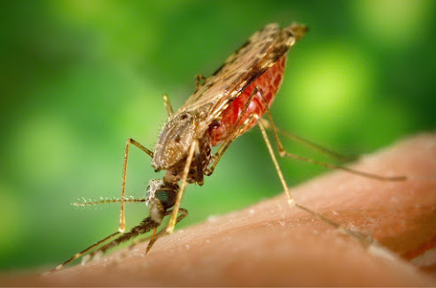
Climate Change Portends Wider Malaria Risk as Mosquitos Spread South and to Higher Elevations in Africa

Anopheles funestus, one of the common mosquito species that transmit malaria in Africa.
Photo Credit: Oberholster Venita

Based on data that span the past 120 years, scientists at Georgetown University Medical Center have found that the mosquitoes responsible for transmitting malaria in Africa are spreading deeper into southern Africa and to higher elevations than previously recorded. The researchers estimate that Anopheles mosquito populations in sub-Saharan Africa have gained an average of 6.5 meters (21 feet) of elevation per year, and the southern limits of their ranges moved south of the equator by 4.7 kilometers (nearly 3 miles) per year.

The study appeared February 15, 2023, in Biology Letters.

“This is exactly what we would expect to see if climate change is helping these species reach colder parts of the continent,” says Colin Carlson, PhD, an assistant research professor at the Center for Global Health Science and Security at Georgetown University Medical Center and lead author of the study. “If mosquitoes are spreading into these areas for the first time, it might help explain some recent changes in malaria transmission that have otherwise been hard to trace back to climate.”

AI with infrared imaging enables precise colon cancer diagnostics

Klaus Gerwert, Stephanie Schörner and Frederik Großerüschkamp (from left) want to improve the diagnosis of colon cancer with the help of artificial intelligence.
Photo Credit: © RUB, Marquard

Artificial intelligence and infrared imaging automatically classify tumors and are faster than previous methods.

The immense progress in the area of therapy options over the past few years has significantly improved the chances of recovery for patients with colon cancer. However, these new approaches, such as immunotherapy, require a precise diagnosis so that they can be tailored to the respective person. Researchers at the Center for Protein Diagnostics PRODI at the Ruhr University Bochum use artificial intelligence in combination with infrared imaging to optimally coordinate the therapy of colon cancer with the individual patient. The label-free and automatable method can complement existing pathological analyzes. The team around Prof. Dr. Klaus Gerwert reports in the journal "European Journal of Cancer" on January 28, 2023.

Deep insights into human tissue within an hour

The PRODI team has been developing a new method of digital imaging for several years: The so-called label-free infrared (IR) imaging measures the genomic and proteomic composition of the tissue examined, i.e. provides molecular information based on the infrared spectra. This information is decoded using artificial intelligence and displayed as false color images. For this purpose, the researchers use image analysis methods from the field of deep learning.

Tuesday, February 14, 2023

Heart failure places a great strain on healthcare

Anna Norhammar, adjunct professor at KI.
Photo Credit: Ulf Sirborn

Patients with heart failure often suffer from co-morbidities, which places a great strain on the healthcare services, a multinational study published in Heart reports. The researchers, who are based at Karolinska Institutet, identify an urgent need to improve risk management of the disease.

Up to 64 million people around the world have heart failure a figure that is expected to rise as populations age and diagnostic methods improve.

According to the new study, there are no multinational studies describing heart failure patients and the consequences of the disease.   

“Given that we know that the incidence of heart failure increases with population age, a modern, broad view of what the heart failure population looks like, involving risks and costs, is important for all forms of care planning,” says Anna Norhammar, adjunct professor at the Cardiology Unit, Department of Medicine (Solna), Karolinska Institutet.

Monday, February 13, 2023

Lung cancer study finds new target for treatment resistance after EGFR inhibitors

Monique Nilsson, Ph.D. | John Heymach, M.D., Ph.D.
Photo Credit: Courtesy of University of Texas M. D. Anderson Cancer Center

Researchers at The University of Texas MD Anderson Cancer Center have identified CD70 as being highly expressed on drug-resistant cancer cells in EGFR-mutant non-small cell lung cancer (NSCLC), highlighting a novel therapeutic target that could be used to eliminate resistant cells remaining after treatment with commonly used EGFR tyrosine kinase inhibitors (TKIs). The study published today in Cancer Cell.

The preclinical research was led by Monique Nilsson, Ph.D., and corresponding author John Heymach, M.D., Ph.D., chair of Thoracic/Head and Neck Medical Oncology. The researchers discovered that CD70, a cell surface protein normally found on immune cells, is highly overexpressed in resistant cells as well as in the residual cancer cells immediately following TKI treatment. They demonstrated that CD70 can be effectively used to target these cells with antibody-drug conjugates (ADCs) or cell therapies in laboratory models.

“Residual cancer cells left over from TKI treatment are essentially a reservoir from which future resistant cells eventually grow,” Heymach said. “These findings set the stage for a really promising approach in which we may give initial effective therapies and immediately follow them with these CD70-targeting drugs to eliminate the remaining residual cells.”

Ingestible sensor could help doctors pinpoint GI difficulties

MIT engineers have shown that they can use magnetic fields to track the location of this ingestible sensor within the GI tract.
Photo Credit: Courtesy of the researchers / Massachusetts Institute of Technology

Engineers at MIT and Caltech have demonstrated an ingestible sensor whose location can be monitored as it moves through the digestive tract, an advance that could help doctors more easily diagnose gastrointestinal motility disorders such as constipation, gastroesophageal reflux disease, and gastroparesis.

The tiny sensor works by detecting a magnetic field produced by an electromagnetic coil located outside the body. The strength of the field varies with distance from the coil, so the sensor’s position can be calculated based on its measurement of the magnetic field.

In the new study, the researchers showed that they could use this technology to track the sensor as it moved through the digestive tract of large animals. Such a device could offer an alternative to more invasive procedures, such as endoscopy, that are currently used to diagnose motility disorders.

“Many people around the world suffer from GI dysmotility or poor motility, and having the ability to monitor GI motility without having to go into a hospital is important to really understand what is happening to a patient,” says Giovanni Traverso, an associate professor of mechanical engineering at MIT and a gastroenterologist at Brigham and Women’s Hospital.

'Natural killer' immune cells can modify tissue inflammation

Professor Mariapia Degli-Esposti and Dr Iona Schuster
Photo Credit: Courtesy of Monash University

Melbourne researchers have improved our understanding of how the immune system is regulated to prevent disease, identifying a previously unknown role of ‘natural killer’ (NK) immune cells.

The Monash University-led study identified a new group of immune cells, known as tissue-resident memory natural killer (NKRM) cells. NKRM cells limited immune responses in tissues and prevented autoimmunity, which is when the immune system makes a mistake and attacks the body's own tissues or organs.

While additional research is required, the discovery may ultimately be used to treat autoimmune diseases like Sjogren’s Syndrome and possibly chronic inflammatory conditions.

Published in Immunity, the preclinical research is led by senior author Professor Mariapia Degli-Esposti and first author Dr Iona Schuster from the Monash Biomedicine Discovery Institute (BDI), in close ongoing collaboration with the Lions Eye Institute.

Saturday, February 11, 2023

Extracts from two wild plants inhibit COVID-19 virus

 Emory University graduate student Caitlin Risener, first author of the study, gathers tall goldenrod in South Georgia.
Photo Credit: Tharanga Samarakoon

Two common wild plants contain extracts that inhibit the ability of the virus that causes COVID-19 to infect living cells, an Emory University study finds. Scientific Reports published the results — the first major screening of botanical extracts to search for potency against the SARS-CoV-2 virus.

In laboratory dish tests, extracts from the flowers of tall goldenrod (Solidago altissima) and the rhizomes of the eagle fern (Pteridium aquilinum) each blocked SARS-CoV-2 from entering human cells.

The active compounds are only present in miniscule quantities in the plants. It would be ineffective, and potentially dangerous, for people to attempt to treat themselves with them, the researchers stress. In fact, the eagle fern is known to be toxic, they warn.

“It’s very early in the process, but we’re working to identify, isolate and scale up the molecules from the extracts that showed activity against the virus,” says Cassandra Quave, senior author of the study and associate professor in Emory School of Medicine’s Department of Dermatology and the Center for the Study of Human Health. “Once we have isolated the active ingredients, we plan to further test for their safety and for their long-range potential as medicines against COVID-19.”

Single drug injection wards off COVID-19 hospitalizations

A single injection of PEG-lambda interferon proved to be effective against all variants of the coronavirus tested by researchers at Stanford Medicine.
Image Credit: Gerd Altmann

In an international, multicenter, pivotal Phase 3 trial, a single under-the-skin injection of a biological drug given to patients within seven days of the onset of COVID-19 symptoms cut the likelihood they needed to be hospitalized in half. Patients treated within three days of showing symptoms fared even better. Among unvaccinated patients who were treated soon after symptom onset, hospitalization likelihood plummeted markedly.

The drug, pegylated lambda-interferon, or PEG-lambda, proved effective against all COVID-19 viral variants tested, including omicron. Side effects were no greater than those reported by placebo recipients.

A report on the success of the randomized, double-blind, placebo-controlled trial of nearly 2,000 newly infected COVID-19 patients was published online Feb. 9 in the New England Journal of Medicine.

PEG-lambda is a synthetic version of lambda-interferon, a naturally occurring protein that infected cells secrete as a first line of defense against viral infection.

Friday, February 10, 2023

Creating 3D objects with sound

The use of sound waves to create a pressure field to print particles. 
Image Credit: © MPI for Medical Research, Heidelberg University/ Kai Melde

Creating 3D objects with sound

Scientists from the Max Planck Institute for Medical Research and the Heidelberg University have created a new technology to assemble matter in 3D. Their concept uses multiple acoustic holograms to generate pressure fields with which solid particles, gel beads and even biological cells can be printed. These results pave the way for novel 3D cell culture techniques with applications in biomedical engineering.

Additive manufacturing or 3D printing enables the fabrication of complex parts from functional or biological materials. Conventional 3D printing can be a slow process, where objects are constructed one line or one layer at a time. Researchers in Heidelberg and Tübingen now demonstrate how to form a 3D object from smaller building blocks in just a single step. “We were able to assemble microparticles into a three-dimensional object within a single shot using shaped ultrasound”, says Kai Melde, postdoc in the group and first author of the study. “This can be very useful for bioprinting. The cells used there are particularly sensitive to the environment during the process”, adds Peer Fischer, Professor at Heidelberg University.

Thursday, February 9, 2023

Disrupted flow of brain fluid may underlie neurodevelopmental disorders

The addition of a magenta tracer molecule illustrates the flow of fluid around the brain, revealing that neurons in the hippocampus (cyan), the brain’s memory center, are awash in fluid. Researchers at Washington University School of Medicine in St. Louis have discovered that this fluid flows to areas critical for normal brain development and function, suggesting that disruptions to its circulation may play an underrecognized role in neurodevelopmental disorders.
Photo Credit: Shelei Pan and Peter Yang/School of Medicine

The brain floats in a sea of fluid that cushions it against injury, supplies it with nutrients and carries away waste. Disruptions to the normal ebb and flow of the fluid have been linked to neurological conditions including Alzheimer’s disease and hydrocephalus, a disorder involving excess fluid around the brain.

Researchers at Washington University School of Medicine in St. Louis created a new technique for tracking circulation patterns of fluid through the brain and discovered, in rodents, that it flows to areas critical for normal brain development and function. Further, the scientists found that circulation appears abnormal in young rats with hydrocephalus, a condition associated with cognitive deficits in children.

The findings, available online in Nature Communications, suggest that the fluid that bathes the brain — known as cerebrospinal fluid — may play an underrecognized role in normal brain development and neurodevelopmental disorders.

Inhalable ‘SHIELD’ Protects Lungs Against COVID-19, Flu Viruses

Photo Credit: Robina Weermeijer

Researchers have developed an inhalable powder that could protect lungs and airways from viral invasion by reinforcing the body’s own mucosal layer. The powder, called Spherical Hydrogel Inhalation for Enhanced Lung Defense, or SHIELD, reduced infection in both mouse and non-human primate models over a 24-hour period, and can be taken repeatedly without affecting normal lung function.

“The idea behind this work is simple – viruses have to penetrate the mucus in order to reach and infect the cells, so we’ve created an inhalable bioadhesive that combines with your own mucus to prevent viruses from getting to your lung cells,” says Ke Cheng, corresponding author of the paper describing the work. “Mucus is the body’s natural hydrogel barrier; we are just enhancing that barrier.”

Cheng is the Randall B. Terry, Jr. Distinguished Professor in Regenerative Medicine at North Carolina State University’s College of Veterinary Medicine and a professor in the NC State/UNC-Chapel Hill Joint Department of Biomedical Engineering.

The inhalable powder microparticles are composed of gelatin and poly(acrylic acid) grafted with a non-toxic ester. When introduced to a moist environment – such as the respiratory tract and lungs – the microparticles swell and adhere to the mucosal layer, increasing the “stickiness” of the mucus.

Large-scale genetic analysis shows microRNAs in human pancreas associated with diabetes

 NIH study identifies new molecules involved in diabetes.
Illustration Credit: CFVI

In a new large-scale genetic analysis, scientists have found a set of small RNA molecules, called microRNAs, in human pancreatic cells that are strongly associated with type 2 diabetes. Researchers discovered the microRNAs in groups of cells called pancreatic islets, which produce hormones, such as insulin, that the body uses to regulate energy levels.

In people with diabetes, the islets fail to produce sufficient insulin to control blood sugar, which is why understanding the basic biology of pancreatic islets is important for human health.

The study, led in part by scientists at the National Human Genome Research Institute (NHGRI), part of the National Institutes of Health, will inform future studies on the early detection and treatment of diabetes. The results were published in Proceedings of the National Academy of the Sciences.

Previous research with animal or cell-based models over the past two decades suggests that certain microRNAs, which are involved in controlling which genes are turned on and off in cells, may help pancreatic islets normally develop and function.

Harmful Effects of Long-Term Alcohol Use Documented in Blood Protein Snapshot

Jon Jacobs recently found that a particular combination of blood proteins indicates alcohol-associated hepatitis, a deadly liver disease. 
Photo Credit: Eddie Pablo III | Pacific Northwest National Laboratory

Biochemist Jon Jacobs has analyzed the blood of patients with diseases and conditions such as Ebola, cancer, tuberculosis, hepatitis, diabetes, Lyme disease, brain injury and influenza.

But never has he seen blood chemistry gone so awry as when he and colleagues took an in-depth look at the protein activity in the blood of patients with alcohol-associated hepatitis, a severe form of liver disease caused by heavy drinking for many years.

“The proteins in these patients are more dysregulated than in any other blood plasma that we’ve analyzed,” said Jacobs, a scientist at the Department of Energy’s Pacific Northwest National Laboratory. “Almost two-thirds of the proteins we measured are at unusual levels. This is a snapshot of what’s going on in the body of a person with this disease and reflects just how severe a disease this is.”

That “snapshot” is a measurement of proteins that change in patients with the disease. The unique combination of changes in protein activity marks an important step toward development of a simple blood test to diagnose alcohol-associated hepatitis.

Jacobs and colleagues, including scientists and physicians from the Veteran Affairs Long Beach Healthcare System and the University of Pittsburgh, published their findings recently in the American Journal of Pathology. Corresponding authors of the study are Jacobs and Timothy Morgan, a gastroenterologist at VA Long Beach who has treated patients with the disease for more than 35 years.

Featured Article

Autism and ADHD are linked to disturbed gut flora very early in life

The researchers have found links between the gut flora in babies first year of life and future diagnoses. Photo Credit:  Cheryl Holt Disturb...

Top Viewed Articles