 |
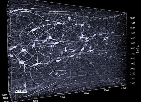
| Three-dimensional image of noradrenergic nerve cells in the envelope of locus coeruleus. Photo credit: Gilvesy et al. |
With the help of a new imaging technique for 3D, researchers at Karolinska Institutet, among others, have been able to characterize a part of the brain that shows the most accumulation of tau protein, an important biomarker for the development of Alzheimer's disease. The results published in the journal Acta Neuropathologica may in the future make it possible to have a more accurate neuropathological diagnosis of Alzheimer's disease spectrum at a very early stage.
Intracellular accumulation of pathological tau protein in the brain is a hallmark of several age-related neurodegenerative diseases, including Alzheimer's disease, which accounts for 60-80 percent of all dementia cases worldwide.
In a new study, researchers at Karolinska Institutet, SciLifeLab in Stockholm and several universities from Hungary, Canada, Germany and France have applied a state-of-the-art immune imaging technology, in combination with light sheet microscopy, to investigate a human brain stem core, locus coeruleus, which is an important core in the mammalian brain.
Early stages of Alzheimer's disease
 |
| Csaba Adori, a researcher at the Department of Neuroscience Source: Karolinska Institutet |
Using 3D imaging of human tissue from the deceased, researchers discovered an exciting complexity and possibly unspecified cellular forms of tau pathology in this brain region as early as the early stages of Alzheimer's disease.
Our study shows that a gradual shrinkage (atrophy) of the dendrite projections is the first morphological sign of degeneration of tau-bearing locus coeruleus nerve cells, ie even before axon damage. Dendrites are the structure that receives impulses from other nerve cells and thus are crucial for communication in the brain. Degeneration of dendrites can lead to serious functional disorders such as overactive nerve cells. This can contribute to severe symptoms that preceded the onset of Alzheimer's disease, including sleep disorders, anxiety and depression, all of which may be related to a distorted function of locus coeruleus, the study's latest author says Csaba Adori, researchers at Department of Neuroscience, Karolinska Institutet.
Supports previous in vitro discoveries
3D analysis detected a non-random distribution of tau-bearing locus coeruleus nerve cells with a cluster tendency. The researchers also discovered that the dendrites of adjacent, clustered tau-bearing nerve cells often showed dendro-dendric contacts. This is in line with the theory that pathological tau can be spread via neuronal projections from one nerve cell to another, as earlier in vitro studies and animal experiments suggest.
In addition, the researchers show that tau pathology is more prominent in a part of the locus coeruleus that projects to the regions of the anterior brain most severely affected by Alzheimer's disease.
The results may represent a clear advance in understanding how brain cell architecture can have an impact on the development and spread of pathological tau protein.
Our results contribute to further understanding of why some brain regions are more affected by Alzheimer's disease than others. In addition, the new methodology applied to human tissue opens the way for more accurate neuropathological diagnostic procedures, even in very early stages of Alzheimer's disease spectrum. This will hopefully contribute to developing more effective prevention strategies for preventing Alzheimer's disease in the future, says Csaba Adori.
The study was supported by the Brain Fund, Olle Engkvist Foundation, Åhlen Foundation, Dementia Association, Lars Hiertas Minne Foundation, Karolinska Institutet, Knut and Alice Wallenberg Foundation, Swedish Research Council, Rossy Family Foundation, Edmond J. Safra Philanthropic Foundation, and Hungarian National Brain Research Program.
Visit Csaba Adori's website for more information and 3D renderings of his research.
Source/Credit: Karolinska Institutet
ns083122_01


.jpg)